Japanese
English
- 有料閲覧
- Abstract 文献概要
- 1ページ目 Look Inside
I.緒言
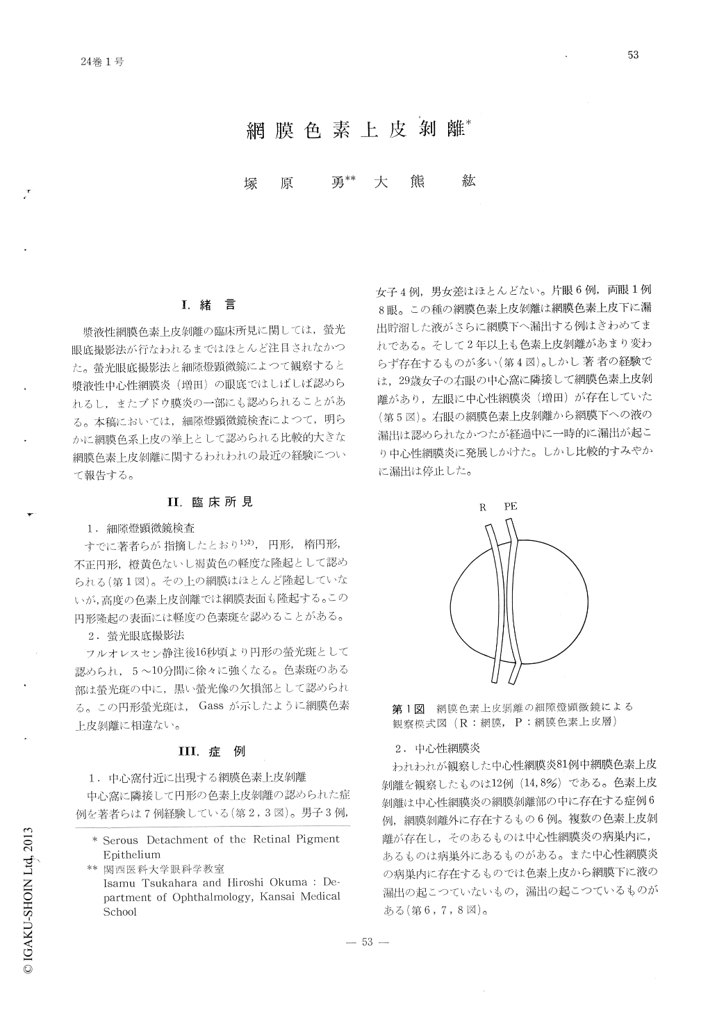
漿液性網膜色素上皮剥離の臨床所見に関しては,螢光眼底撮影法が行なわれるまではほとんど注目されなかつた。螢光眼底撮影法と細隙燈顕微鏡によつて観察すると漿液性中心性網膜炎(増田)の眼底ではしばしば認められるし,またブドウ膜炎の一部にも認められることがある。本稿においては,細隙燈顕微鏡検査によつて,明らかに網膜色系上皮の挙上として認められる比較的大きな網膜色素上皮剥離に関するわれわれの最近の経験について報告する。
This report concerns larger lesion of serousdetachment of the pigment epithelium which is seen as a well difined round or oval shallow elevation of the pigment epithelium by slit lamp microscopy and demonstrated as a focal area of diffuse fluorescence by fluorescence fundus angiography.
Isolated parafoveal lesion was observed in 7 cases and similar lesions were found in 12 out of 81 cases of Retinitis centralis (central serous choroidopathy).In 2 cases of uveitis which showed positive toxoplasmic hemagglutinin test deta-ched pigment epithelium was observed.

Copyright © 1970, Igaku-Shoin Ltd. All rights reserved.


