Japanese
English
- 有料閲覧
PPV購入案内
1,320円 (1,200円+税10%) こちらは、407ページから411ページの文献です。購入には医書.jp本会員登録が必要です。
会員登録完了後に改めて上記「購入サイトへ」を選択してください。
または「購入サイトへ」選択後に「購入ログイン」、「新規会員登録」の順に進んでください。
会員登録について詳しくはをご確認ください。
- Abstract 文献概要
- 1ページ目 Look Inside
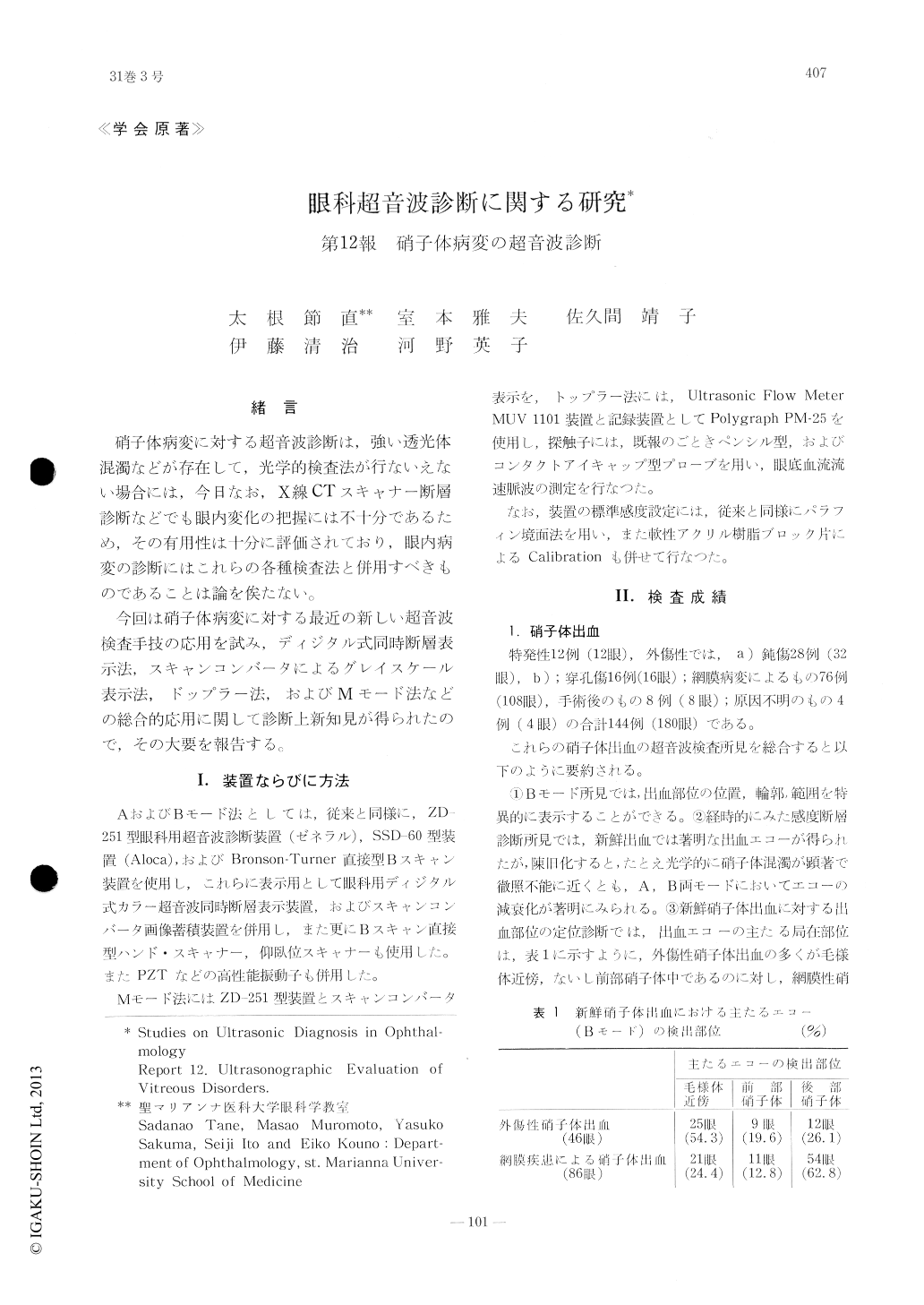
緒 言
硝子体病変に対する超音波診断は,強い透光体混濁などが存在して,光学的検査法が行ないえない場合には,今日なお,X線CTスキャナー断層診断などでも眼内変化の把握には不十分であるため,その有用性は十分に評価されており,眼内病変の診断にはこれらの各種検査法と併用すべきものであることは論を俟たない。
今回は硝子体病変に対する最近の新しい超音波検査手技の応用を試み,ディジタル式同時断層表示法,スキャンコンバータによるグレイスケール表示法,ドップラー法,およびMモード法などの総合的応用に関して診断上新知見が得られたので,その大要を報告する。
Characteristics of vitreous disorders can be depicted most vividly with A and B-scan ultra-sonography. Analysis of the location, density, and complication of the vitreous hemorrhage, opacities, foreign body, proliferative change, and retinal detachment may help in predicting the extent of visual recovery. In addition, ac-curate knowledge of the pattern of the hemor-rhage and other ocular abnormalities enables more adequate preoperative evaluation for vit-reous surgery or retinal repair.
Copyright © 1977, Igaku-Shoin Ltd. All rights reserved.