Japanese
English
- 有料閲覧
PPV購入案内
1,320円 (1,200円+税10%) こちらは、55ページから63ページの文献です。購入には医書.jp本会員登録が必要です。
会員登録完了後に改めて上記「購入サイトへ」を選択してください。
または「購入サイトへ」選択後に「購入ログイン」、「新規会員登録」の順に進んでください。
会員登録について詳しくはをご確認ください。
- Abstract 文献概要
- 1ページ目 Look Inside
緒言
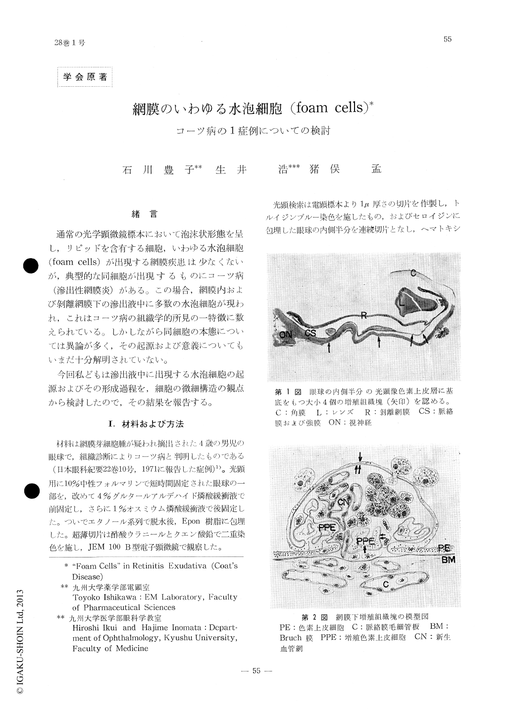
通常の光学顕微鏡標本において泡沫状形態を呈し,リピッドを含有する細胞,いわゆる水泡細胞(foam cells)が出現する網膜疾患は少なくないが,典型的な同細胞が出現するものにコーツ病(滲出性網膜炎)がある。この場合,網膜内および剥離網膜下の滲出液中に多数の水泡細胞が現われ,これはコーツ病の組織学的所見の一特徴に数えられている。しかしながら同細胞の本態については異論が多く,その起源および意義についてもいまだ十分解明されていない。
今回私どもは滲出液中に出現する水泡細胞の起源およびその形成過程を,細胞の微細構造の観点から検討したので,その結果を報告する。
Of a variety of histological pictures of Coats' disease, one of the characteristic findings is the presence of aggregates of bladder-like, lipid-laden cells ("foam cells") in the retina and in the subretinal exudate. Opinion is divided on the origin and significance of the cells.
An enucleated eyeball of 4-year-old boy with Coats' disease was examined electron microsco-pically. Foam cells are mainly located in the subretinal exudate and in the nodular fibrous tissues formed between the retina and the cho-roid, and resting on the pigment epithelium. A number of foam cells, however, are not found in the retina proper.
Copyright © 1974, Igaku-Shoin Ltd. All rights reserved.