Japanese
English
特集 第26回日本臨床眼科学会講演集(その4)
学会原著
緑内障眼前房隅角の微細構造—(1)走査宣顕的研究
The Fine Structure of the Iridocorneal Angle Tissue of Glaucomatous Eyes (1) A Scanning Electron Microscopic Study
瀬川 雄三
1
Katsuzo Segawa
1
1信州大学医学部眼科学教室
1Ophthalmological Clinic, Shinshu University
pp.461-463
発行日 1973年4月15日
Published Date 1973/4/15
DOI https://doi.org/10.11477/mf.1410204937
- 有料閲覧
PPV購入案内
1,320円 (1,200円+税10%) こちらは、461ページから463ページの文献です。
PPV(ペイ・パー・ビュー)とは、論文(記事)単位で購入・閲覧ができるサービスです。
購入には医書.jp本会員登録が必要です。
会員登録完了後に改めて上記「購入サイトへ」を選択してください。
または「購入サイトへ」選択後に「購入ログイン」、「新規会員登録」の順に進んでください。
会員登録について詳しくはをご確認ください。
購入には医書.jp本会員登録が必要です。
会員登録完了後に改めて上記「購入サイトへ」を選択してください。
または「購入サイトへ」選択後に「購入ログイン」、「新規会員登録」の順に進んでください。
会員登録について詳しくはをご確認ください。
- Abstract 文献概要
- 1ページ目 Look Inside
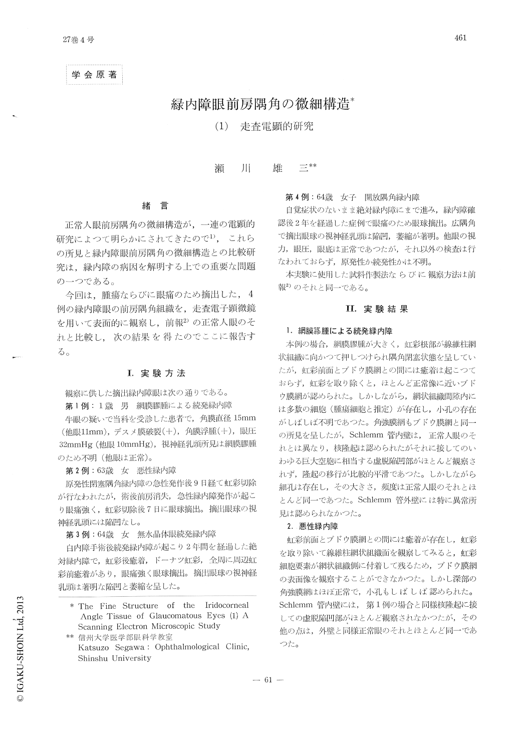
緒言
正常人眼前房隅角の微細構造が,一連の電顕的研究によつて明らかにされてきたので1),これらの所見と緑内障眼前房隅角の微細構造との比較研究は,緑内障の病因を解明する上での重要な問題の一つである。
今回は,腫瘍ならびに眼痛のため摘出した,4例の緑内障眼の前房隅角組織を,走査電子顕微鏡を用いて表面的に観察し,前報2)の正常人眼のそれと比較し,次の結果を得たのでここに報告する。
The iridocorneal angle tissue of four glaucom-atous eyes enucleated because of retinoblastoma or ocular pains, which consisted of secondary angle-closure glaucoma due to retinoblastoma, malignant glaucoma, secondary angle-closure glaucoma due to posterior synechias after lens extraction and open-angle absolute glaucoma, was examined with a scanning electron micro-scope and compared with that of normal eyes2).
(1) In all glaucomatous eyes studied, unlike in normal eyes, few collapsed giant vacuoles were found on the trabecular wall of Schlemm'scanal.
Copyright © 1973, Igaku-Shoin Ltd. All rights reserved.