Japanese
English
- 有料閲覧
PPV購入案内
1,320円 (1,200円+税10%) こちらは、721ページから731ページの文献です。購入には医書.jp本会員登録が必要です。
会員登録完了後に改めて上記「購入サイトへ」を選択してください。
または「購入サイトへ」選択後に「購入ログイン」、「新規会員登録」の順に進んでください。
会員登録について詳しくはをご確認ください。
- Abstract 文献概要
- 1ページ目 Look Inside
I.緒言
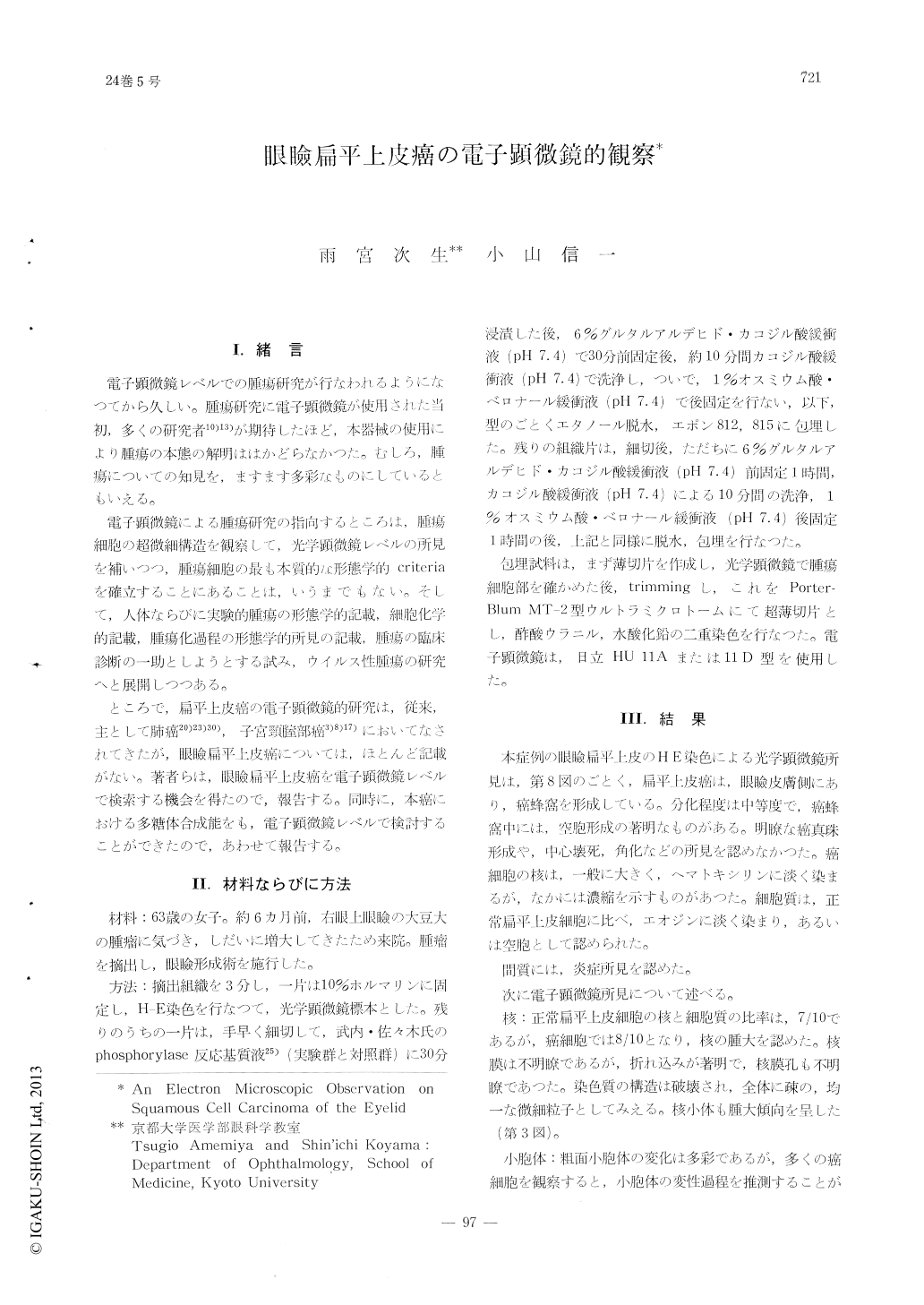
電子顕微鏡レベルでの腫瘍研究が行なわれるようになつてから久しい。腫瘍研究に電子顕微鏡が使用された当初,多くの研究者10)13)が期待したほど,本器械の使用により腫瘍の本態の解明ははかどらなかつた。むしろ,腫瘍についての知見を,ますます多彩なものにしているともいえる。
電子顕微鏡による腫瘍研究の指向するところは,腫瘍細胞の超微細構造を観察して,光学顕微鏡レベルの所見を補いつつ,腫瘍細胞の最も本質的な形態学的criteriaを確立することにあることは,いうまでもない。そして,人体ならびに実験的腫瘍の形態学的記載,細胞化学的記載,腫瘍化過程の形態学的所見の記載,腫瘍の臨床診断の一助としようとする試み,ウイルス性腫瘍の研究へと展開しつつある。
Squamous cell carcinoma of the eyelid was studied by electron microscope.
The nuclei of tumor cells revealed enlarge-ment, increased nuclear membrane infoldings and enlargement of the nucleoli.
In the cytoplasm, mitochondria decreased in number, were enlarged and swollen. Glycogen was few or absent. The endoplasmic reticulum showed prominent vacuolation, loss of tonofila-ments and increase of free-ribosomes. The des-mosomes either decreased in number or were lost. There was an absence of the usual tono-fibrils at the desmosomes. The basement mem-brane was frequently present in imperfect form. Where the basement membrane was absent, small protrusion of carcinoma cell cytoplasm was seen in the adjacent stroma.
Copyright © 1970, Igaku-Shoin Ltd. All rights reserved.