Japanese
English
- 有料閲覧
PPV購入案内
1,320円 (1,200円+税10%) こちらは、1485ページから1488ページの文献です。購入には医書.jp本会員登録が必要です。
会員登録完了後に改めて上記「購入サイトへ」を選択してください。
または「購入サイトへ」選択後に「購入ログイン」、「新規会員登録」の順に進んでください。
会員登録について詳しくはをご確認ください。
- Abstract 文献概要
- 1ページ目 Look Inside
- 参考文献 Reference
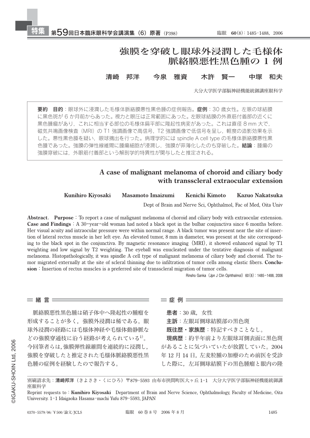
目的:眼球外に浸潤した毛様体脈絡膜悪性黒色腫の症例報告。症例:30歳女性。左眼の球結膜に黒色斑が6か月前からあった。視力と眼圧は正常範囲にあった。左眼球結膜の外直筋付着部の近くに黒色腫瘤があり,これに相当する部位の毛様体【扁】平部に隆起性病変があった。これは直径8mm大で,磁気共鳴画像検査(MRI)のT1強調画像で高信号,T2強調画像で低信号を呈し,軽度の造影効果を示した。悪性黒色腫を疑い,眼球摘出を行った。病理学的にはspindle A cell typeの毛様体脈絡膜悪性黒色腫であった。強膜の弾性線維間に腫瘍細胞が浸潤し,強膜が菲薄化したのち穿破した。結論:腫瘍の強膜穿破には,外眼筋付着部という解剖学的特異性が関与したと推定される。
Purpose:To report a case of malignant melanoma of choroid and ciliary body with extraocular extension. Case and Findings:A 30-year-old woman had noted a black spot in the bulbar conjunctiva since 6 months before. Her visual acuity and intraocular pressure were within normal range. A black tumor was present near the site of insertion of lateral rectus muscle in her left eye. An elevated tumor, 8mm in diameter, was present at the site corresponding to the black spot in the conjunctiva. By magnetic resonance imaging(MRI), it showed enhanced signal by T1 weighting and low signal by T2 weighting. The eyeball was enucleated under the tentative diagnosis of malignant melanoma. Histopathologically, it was spindle A cell type of malignant melanoma of ciliary body and choroid. The tumor migrated externally at the site of scleral thinning due to infiltration of tumor cells among elastic fibers. Conclusion:Insertion of rectus muscles is a preferred site of transscleral migration of tumor cells.
Rinsho Ganka(Jpn J Clin Ophthalmol)60(8):1485-1488, 2006
Copyright © 2006, Igaku-Shoin Ltd. All rights reserved.