Japanese
English
- 有料閲覧
PPV購入案内
1,320円 (1,200円+税10%) こちらは、1153ページから1156ページの文献です。購入には医書.jp本会員登録が必要です。
会員登録完了後に改めて上記「購入サイトへ」を選択してください。
または「購入サイトへ」選択後に「購入ログイン」、「新規会員登録」の順に進んでください。
会員登録について詳しくはをご確認ください。
- Abstract 文献概要
- 1ページ目 Look Inside
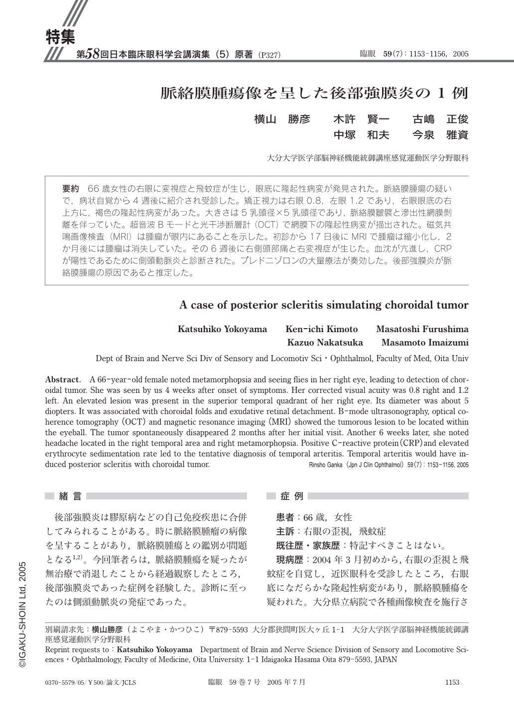
66歳女性の右眼に変視症と飛蚊症が生じ,眼底に隆起性病変が発見された。脈絡膜腫瘍の疑いで,病状自覚から4週後に紹介され受診した。矯正視力は右眼0.8,左眼1.2であり,右眼眼底の右上方に,褐色の隆起性病変があった。大きさは5乳頭径×5乳頭径であり,脈絡膜皺襞と滲出性網膜剝離を伴っていた。超音波Bモードと光干渉断層計(OCT)で網膜下の隆起性病変が描出された。磁気共鳴画像検査(MRI)は腫瘤が眼内にあることを示した。初診から17日後にMRIで腫瘤は縮小化し,2か月後には腫瘤は消失していた。その6週後に右側頭部痛と右変視症が生じた。血沈が亢進し,CRPが陽性であるために側頭動脈炎と診断された。プレドニゾロンの大量療法が奏効した。後部強膜炎が脈絡膜腫瘍の原因であると推定した。
A 66-year-old female noted metamorphopsia and seeing flies in her right eye,leading to detection of choroidal tumor. She was seen by us 4 weeks after onset of symptoms. Her corrected visual acuity was 0.8 right and 1.2 left. An elevated lesion was present in the superior temporal quadrant of her right eye. Its diameter was about 5 diopters. It was associated with choroidal folds and exudative retinal detachment. B-mode ultrasonography,optical coherence tomography(OCT)and magnetic resonance imaging(MRI)showed the tumorous lesion to be located within the eyeball. The tumor spontaneously disappeared 2 months after her initial visit. Another 6 weeks later,she noted headache located in the right temporal area and right metamorphopsia. Positive C-reactive protein(CRP)and elevated erythrocyte sedimentation rate led to the tentative diagnosis of temporal arteritis. Temporal arteritis would have induced posterior scleritis with choroidal tumor.
Copyright © 2005, Igaku-Shoin Ltd. All rights reserved.