Japanese
English
- 有料閲覧
PPV購入案内
1,320円 (1,200円+税10%) こちらは、1135ページから1139ページの文献です。購入には医書.jp本会員登録が必要です。
会員登録完了後に改めて上記「購入サイトへ」を選択してください。
または「購入サイトへ」選択後に「購入ログイン」、「新規会員登録」の順に進んでください。
会員登録について詳しくはをご確認ください。
- Abstract 文献概要
- 1ページ目 Look Inside
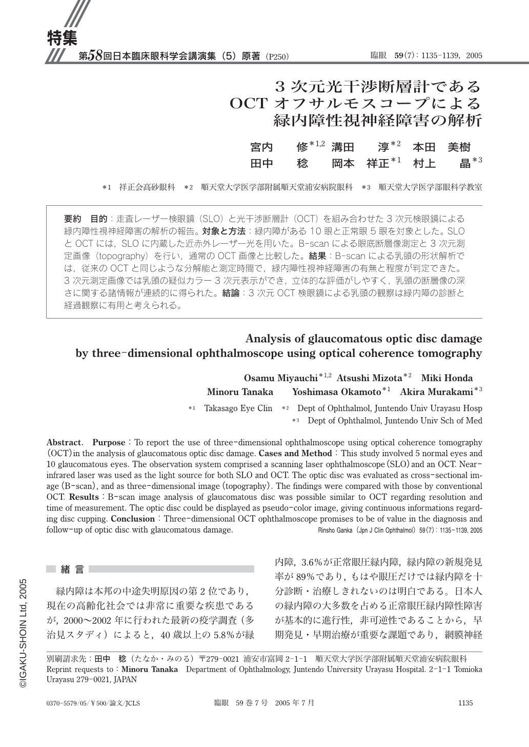
目的:走査レーザー検眼鏡(SLO)と光干渉断層計(OCT)を組み合わせた3次元検眼鏡による緑内障性視神経障害の解析の報告。対象と方法:緑内障がある10眼と正常眼5眼を対象とした。SLOとOCTには,SLOに内蔵した近赤外レーザー光を用いた。B-scanによる眼底断層像測定と3次元測定画像(topography)を行い,通常のOCT画像と比較した。結果:B-scanによる乳頭の形状解析では,従来のOCTと同じような分解能と測定時間で,緑内障性視神経障害の有無と程度が判定できた。3次元測定画像では乳頭の疑似カラー3次元表示ができ,立体的な評価がしやすく,乳頭の断層像の深さに関する諸情報が連続的に得られた。結論:3次元OCT検眼鏡による乳頭の観察は緑内障の診断と経過観察に有用と考えられる。
Purpose:To report the use of three-dimensional ophthalmoscope using optical coherence tomography(OCT)in the analysis of glaucomatous optic disc damage. Cases and Method:This study involved 5 normal eyes and 10 glaucomatous eyes. The observation system comprised a scanning laser ophthalmoscope(SLO)and an OCT. Near-infrared laser was used as the light source for both SLO and OCT. The optic disc was evaluated as cross-sectional image(B-scan),and as three-dimensional image(topography). The findings were compared with those by conventional OCT. Results:B-scan image analysis of glaucomatous disc was possible similar to OCT regarding resolution and time of measurement. The optic disc could be displayed as pseudo-color image,giving continuous informations regarding disc cupping. Conclusion:Three-dimensional OCT ophthalmoscope promises to be of value in the diagnosis and follow-up of optic disc with glaucomatous damage.
Copyright © 2005, Igaku-Shoin Ltd. All rights reserved.