Japanese
English
- 有料閲覧
PPV購入案内
1,320円 (1,200円+税10%) こちらは、383ページから387ページの文献です。購入には医書.jp本会員登録が必要です。
会員登録完了後に改めて上記「購入サイトへ」を選択してください。
または「購入サイトへ」選択後に「購入ログイン」、「新規会員登録」の順に進んでください。
会員登録について詳しくはをご確認ください。
- Abstract 文献概要
- 1ページ目 Look Inside
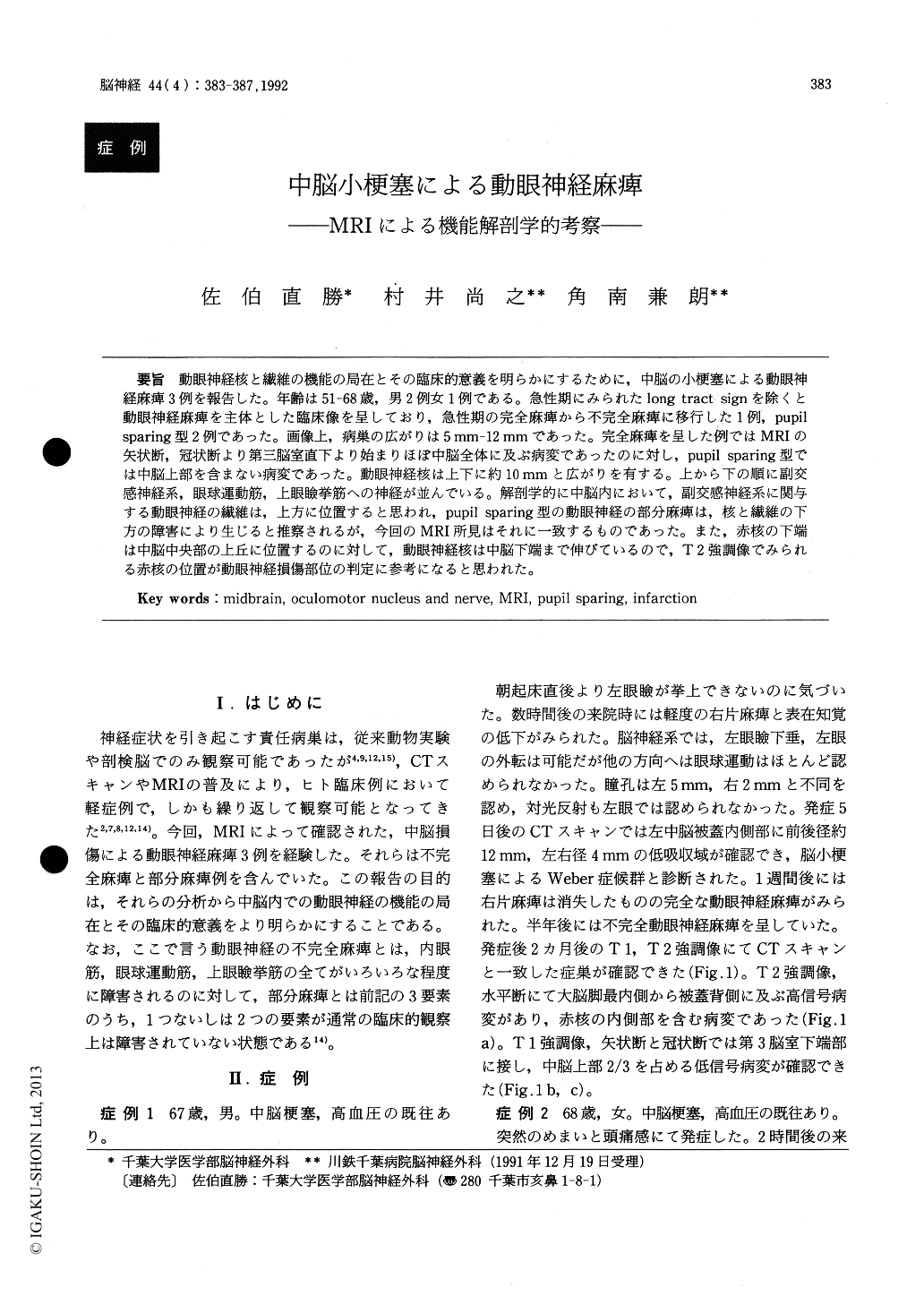
動眼神経核と繊維の機能の局在とその臨床的意義を明らかにするために,中脳の小梗塞による動眼神経麻痺3例を報告した。年齢は51-68歳,男2例女1例である。急性期にみられたlong tract signを除くと動眼神経麻痺を主体とした臨床像を呈しており,急性期の完全麻痺から不完全麻痺に移行した1例,pupilsparing型2例であった。画像上,病巣の広がりは5mm−12mmであった。完全麻痺を呈した例ではMRIの矢状断,冠状断より第三脳室直下より始まりほぼ中脳全体に及ぶ病変であったのに対し,pupil sparing型では中脳上部を含まない病変であった。動眼神経核は上下に約10mmと広がりを有する。上から下の順に副交感神経系,眼球運動筋,上眼瞼挙筋への神経が並んでいる。解剖学的に中脳内において,副交感神経系に関与する動眼神経の繊維は,上方に位置すると思われ,pupil sparing型の動眼神経の部分麻痺は,核と繊維の下方の障害により生じると推察されるが,今回のMRI所見はそれに一致するものであった。また,赤核の下端は中脳中央部の上丘に位置するのに対して,動眼神経核は中脳下端まで伸びているので,T2強調像でみられる赤核の位置が動眼神経損傷部位の判定に参考になると思われた。
This is a report of 3 cases presented with oculomotor nerve palsy caused by small midbrain infarct. The aim of this report is to clarify the functional topography of intranuclear and intrafas-cicular portion of the oculomotor nerve with MRI. Three cases are 2 males and 1 female, ranging 51 to 68 years in age. Except for the long tract signs at the acute stage, cardinal sings were all eye-related, incomplete in 1 case and pupil sparing-type in 2 cases. In MRI, the size of the lesion extended 5 to 12 mm. In the incomplete palsy case, the infarction extended from the level immediately below the 3rd ventricle into the whole length of midbrain, whereas in the pupil-sparing types, more limited lesion ex-cluding the upper part of the midbrain was noted. Anatomically the longitudinal size of the nucleus is 10mm and nerves functionally related to pupil reac-tion, eye motion and eyelid elevation are arranged in rosrocaudal order. Therefore, it is speculated that in midbrain, intrafascicular location of nerve fibers associated with pupil reaction is rostral and oculomotor nerve palsy of pupil sparing type is caused by the lesion excluding the rostral midbrain. MRI findings of the present 3 cases are compatible with this speculation.
The lowest border of red nucleus is at the level of superior colliculus, whereas oculomotor nucleus has its lowest margin at the inferior colliculus. There-fore, red nuleus becomes an informative landmark to visualized the level of oculomotor nerve injury, since the red nucleus is clearly demonstrated in high intensity in T2 weighted image.
Copyright © 1992, Igaku-Shoin Ltd. All rights reserved.