Japanese
English
- 有料閲覧
- Abstract 文献概要
- 1ページ目 Look Inside
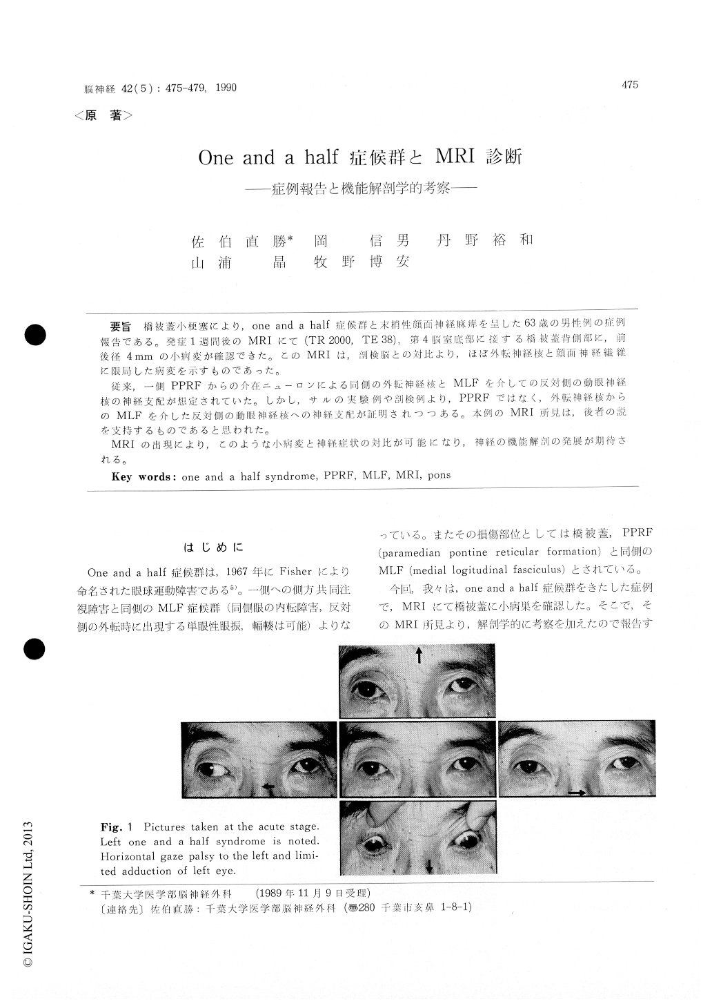
橋被蓋小梗塞により,one and a half症候群と末梢性顔面神経麻痺を呈した63歳の男性症例報告である。発症1週間後のMRIにて(TR 2000, TE 38),第4脳室底部に接する橋被蓋背側部に,前後径4mmの小病変が確認できた。このMRIは,剖検脳との対比より,ほぼ外転神経顔面神経繊維に限局した病変を示すものであった.
従来,一側PPRFからの介在ニューロンによる同側の外転神経核とMLFを介しての反対側の動眼神経核の神経支配が想定されていた。しかし,サルの実験例や剖検例より,PPRFではなく,外転神経核からのMLFを介した反対側の動眼神経核への神経支配が証明されつつある。本例のMRI所見は、後者の説を支持するものであると思われた。
MRIの出現により,このような小病変と神経症状の対比が可能になり,神経の機能解剖の発展が期待される。
This is a case report of 63 year old men who presented one and a half syndrome with ipsilate-ral peripheral type facial palsy due to lacunar infarct. MRI taken one week after the onset (TR 2000, TE 38) demonstrated small high intensity lesion, 4 mm in diameter, located at the dorsal portion of pontine tegmentum, contacting with the floor of 4 th ventricle. This MRI coincides with the lesion limited to the abducens nucleus and genu of facial nerve. Traditionally, projections from the PPRF to the ipsilateral abducens nucleus and opposite MLF was postulated. Recently, how-ever, experiment on monkey and autopsy cases sho-wed projection from abducens nucleus, instead of PPRF, to the opposite MLF has been proposed. MRI findings in this case support the latter hypo-thesis. It is expected that, with the advent of MRI, more meticulous functional neuroanatomy will be developed.

Copyright © 1990, Igaku-Shoin Ltd. All rights reserved.


