Japanese
English
- 有料閲覧
PPV購入案内
1,320円 (1,200円+税10%) こちらは、241ページから245ページの文献です。購入には医書.jp本会員登録が必要です。
会員登録完了後に改めて上記「購入サイトへ」を選択してください。
または「購入サイトへ」選択後に「購入ログイン」、「新規会員登録」の順に進んでください。
会員登録について詳しくはをご確認ください。
- Abstract 文献概要
- 1ページ目 Look Inside
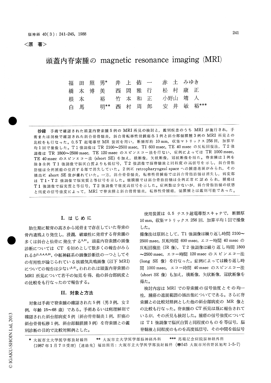
抄録 手術で確認された頭蓋内脊索腫5例のMRI所見の検討と,鑑別疾患のうちMRIが施行され,手術または剖検で確認された斜台骨骨髄炎,斜台骨転移性骨腫瘍各1例と斜台部髄膜腫3例のMRI所見との比較をも行なった。0.5T超電導型MR装置を用い,断層厚約10 mm,収集マトリックス256回,加算平均1回で撮像した。T1強調像はTR 2100〜2500 msec, TI 600 msec, TE 40 msecの反転回復法,T 2強調像はTR 1800〜2500 msec,TE 120 msecのスピンエコー法を行ない,症例によってはTR 1000 msec,TE 40 msecのスピンエコー法(short SE)を加え,横断像,矢状断像,冠状断像を得た。脊索腫は1例を除き全例T1強調像で脳灰白質よりも低信号,T2強調像で脳脊髄液と1同程度の高信号を示し,斜台骨脂肪髄は全例腫瘍の位置する部で消失していた。2例にretropharyngeal spaceへの腫瘍進展がみられ,その描出にshort SE像が優れていた。一方,斜台骨骨髄炎,転移性骨腫瘍では斜台骨脂肪髄は消失し,病変部はT1\T2強調像で脳実質と等信号を示した。髄膜腫では斜台骨脂肪髄は全例正常に認められ,腫瘍はT1強調像で脳実質と等信号,T2強調像で軽度高信号を示した。症例数は少ないが,斜台骨脂肪髄の状態と病変の信号強度によって,MRIで脊索腫と斜台骨骨髄炎,転移性骨腫瘍,髄膜腫とは鑑別可能であった。
MR images of 5 patients with intracranial chor-doma were evaluated and compared with those of other clival lesions (1 clival osteomyelitis, 1 metastatic clival tumor, 3 clival meningiomas). The MR examination was performed using a 0.5 T superconductive magnet, with approxima-tely 10 mm section thickness, one average and a 256×256 matrix. T 1 weighted images were obtainned by inversion recovery (IR) with TR 2100-2500 msec, TI 600 msec and TE 40 msec. T 2 weighted images were obtained by spin echo pulse sequence with TR 1800-2500 msec and TE 120 msec (long SE). In several cases, the spin echo pulse sequences with TR 1000 msec and TE 40 msec (short SE) were also done. Multiplaned images were obtained.
Four of 5 intracranial chordomas were low in intensity compared to cerebral gray matter on T1 weighted images, and all of 5 chordomas were as high in intensity as cerebrospinal fluid or higher than that of cerebrospinal fluid on T2 weighted images. Clival fatty marrow is high intensity on T 1 weighted images. Clival involve-ment by a tumor was a clearly demonstrated as disappearance of this high intensity in all cases. In two cases, the tumor extended to the retro-pharyngeal space and this was detected clearly on short SE image.
Although clival fatty marrow was disappeared, osteomyelitis and metastatic tumor in clivus were iso-intense to cerebral gray matter on both T 1 and T 2 weighted images.
All of 3 clival meningiomas showed iso-inten-sity to cerebral gray matter on T 1 weighted images and slightly high intensity to brain on T 2 weighted images, and clival fatty marrow was normal in all 3 cases.
Although our experiences are limited in num-ber, intracranial chordoma appeared to be diffe-rentiated from other clival lesions.
Copyright © 1988, Igaku-Shoin Ltd. All rights reserved.