Japanese
English
- 有料閲覧
PPV購入案内
1,320円 (1,200円+税10%) こちらは、991ページから996ページの文献です。購入には医書.jp本会員登録が必要です。
会員登録完了後に改めて上記「購入サイトへ」を選択してください。
または「購入サイトへ」選択後に「購入ログイン」、「新規会員登録」の順に進んでください。
会員登録について詳しくはをご確認ください。
- Abstract 文献概要
- 1ページ目 Look Inside
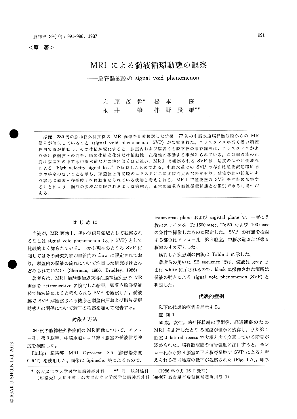
抄録 289例の脳神経外科症例のMR画像を比較検討した結果,77例の中脳水道脳脊髄液腔からのMR信号が消失していること(signal void phenomenon=SVP)が観察された。エラスタンスが高く硬い頭蓋腔内で脳が拍動し,その体積が変化すると,脳室内および脳表くも膜下腔の脳脊髄液は,エラスタンスがより低い脊髄腔との間を,脳の体積変化分だけ拍動性,往復性に移動する事が知られている。この髄液流の速度は脳室系の中でも中脳水道などの狭い部分ほど速い。MRIで観察されるSVPは,速度のはやい髄液流による"high velocity signal loss"を反映したものである。中脳水道でのSVPの存在は髄液流通路に閉塞や狭窄のないことを示し,頭蓋腔と脊髄腔のエラスタンスに比較的大きな差が有り,髄液が脳の拍動により容易に頭蓋—脊髄腔間を移動させられている状態と考えられる。MR Iで髄液腔のSVPを詳細に観察することにより,髄液の脈流が制限されるような病態と,正常の頭蓋内髄液循環状態とを鑑別できる可能性がある。
In a comparative study of MR images of 289 neurosurgical patients, loss of the signal intensity (signal void phenomenon) of CSF in the aqueduct was observed in 77 patients. This signal void phenomenon was seen most frequently in infants with chronic subdural hematoma (12 of 18) andpatients of all age groups suffering from com-municating hydrocephalus (10 of 14).
It is konwn that CSF in the cranial cavity flows toward the spinal CSF space in to and fro manner responding to brain parenchyma pulsations. The velocity of this flow is to be faster in the narrower parts through the ventricular systems such as the aqueduct, Monro's foramen and the 4th ventricles. We think that in T 2 weighted images signal void phenomenon reflects "high velocity signal loss" due to CSF flow. When the subarachnoid adhesions secondary to subarachnoid hemorrhage stagnate CSF flow in the subarachnoid space, the intraven-tricular CSF flow forms the main buffer for changes of the brain volume. This causes an increase in the amplitude of the pulsatile flow in the ven-tricular systems. Therefore the signal void pheno-menon in the aqueductal CSF becomes more pro-nounced.
It may be possible to differentiate normal cir-culation of CSF from abnormal with the bigger amplitude of CSF pulsatile flow, to understand the mechanisms of the normal pressure hydrocephalus or to diagnose a shunt malfunction. Therefore more insight in the CSF flow as imaged by MRI is needed, quantification of CSF flow will be the subjects of our further research.
Copyright © 1987, Igaku-Shoin Ltd. All rights reserved.