Japanese
English
- 有料閲覧
PPV購入案内
1,320円 (1,200円+税10%) こちらは、295ページから302ページの文献です。購入には医書.jp本会員登録が必要です。
会員登録完了後に改めて上記「購入サイトへ」を選択してください。
または「購入サイトへ」選択後に「購入ログイン」、「新規会員登録」の順に進んでください。
会員登録について詳しくはをご確認ください。
- Abstract 文献概要
- 1ページ目 Look Inside
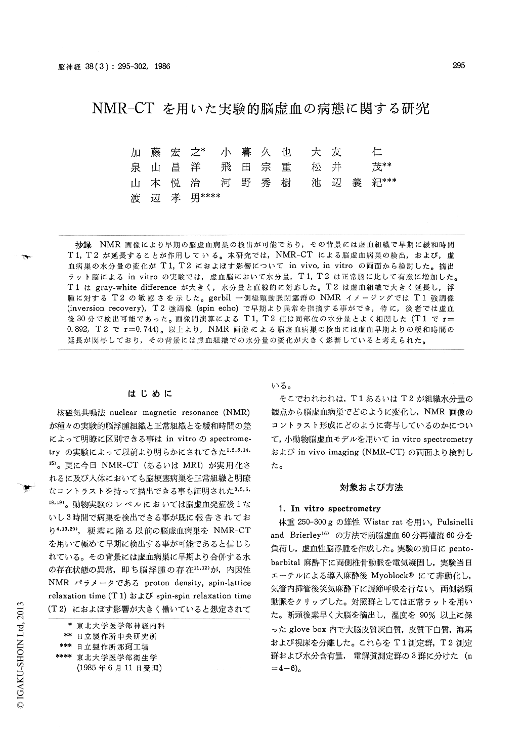
抄録 NMR画像により早期の脳虚血病巣の検出が可能であり,その背景には虚血組織で早期に緩和時間T1,T2が延長することが作用している。本研究では, NMR-CTによる脳虚血病巣の検出,および,虚血病巣の水分量の変化がT1,T2におよぼす影響についてin vivo, in vitroの両面から検討した。摘出ラット脳によるin vitroの実験では,虚血脳において水分量,T1,T2は正常脳に比して有意に増加した。T1はgray・white differenceが大きく,水分量と直線的に対応した。T2は虚血組織で大きく延長し,浮腫に対するT2の敏感さを示した。gerbil一側総頸動脈閉塞群のNMRイメージングではT1強調像(inversio recovery),T2強調像(spin echo)で早期より異常を指摘する事ができ,特に,後者では虚血後30分で検出可能であった。画像間演算によるT1,T2値は同部位の水分量とよく相関した(T1でr=0.892,T2でr=0.744)。以上より, NMR画像による脳虚血病巣の検出には虚血早期よりの緩和時間の延長が関与しており,その背景には虚血組織での水分量の変化が大きく影響していると考えられた。
Recent studies on proton NMR imaging revealed its remarkable sensitivity for detecting cerebral ischemia. Since proton NMR reflects the distribu-tion and state of water in the brain, an NMR imager becomes a sensitive in vivo detector of brain edema developing soon after the energy state is compromized by ischemia. To further cla-rify the usefulness of NMR imaging to characte-rize the ischemia-induced changes, correlations between T 1 and T 2 relaxation times and water content of the normal and ischemic rat and gerbil brain were studied by means of both spectroscopic and in vivo imaging methods.
In the spectroscopic experiment on excised rat brain (cortex, white matter, hippocampus and thalamus for normal and ischemia-laden brain), T 1 and T 2 relaxation times and water content were determined. The ischemic insult was induced for 60 min by the method of Pulsinelli followed by 60 min of reperfusion. All of the T 1, T 2 and water content significantly increased in the ischemic tis-sue. Gray-white difference was evident in T 1 and T 1 was linearly correlated with the water content of the tissue. T 2 was by far prolonged in the isc-hemic tissue compared with the increase in the water content, showing greater sensitivity of T 2 for detection of ischemia.
In the imaging experiment, coronal NMR ima-ging at O.5 tesla was performed employing proton density-weighted saturation recovery (TR =1.6 s, TE= 14 ms), T 1-weighted inversion recovery (TR =1.6 s, TI= 300 ms, TE= 14 ms) and T 2-weighted spin echo (TR =1.6 s, TE= 106 ms) pulse sequences. Spatial resolution of the images was excellent (0.3-0.5 mm) and the imaging of a gerbil brain clearly delineated anatomy separating cortex, whi- to matter, hippocampus and thalamus. Hemispheric ischemia of a gerbil brain was detected as early as 30 min after occlusion of the carotid artery in T 2-weighted images and the evolution of the le-sion was clearly picturized in T 1- and T 2-weight-ed images. On the other hand, SR images were far less sensitive. Calculated T 1 and T 2 relaxa-tion times by the pixel-to-pixel computation indi-cated progress of the lesion and excellently cor-related with the water content of the tissue (r= 0.892 and 0.744 for T 1 and T 2, respectively).
As a result, proton NMR imaging could detect cerebral ischemia as early as 30 min after insult in terms of early alteration of T 1 and T 2 relaxation times in the ischemic tissue. Furthermore, calcula-ted T 1 and T 2 values excellently correlated with the water content and were considered to be the in vivo indices of the dynamic state of water or the degree of the damage in the ischemic tissue.
Copyright © 1986, Igaku-Shoin Ltd. All rights reserved.