Japanese
English
- 有料閲覧
PPV購入案内
1,320円 (1,200円+税10%) こちらは、921ページから926ページの文献です。購入には医書.jp本会員登録が必要です。
会員登録完了後に改めて上記「購入サイトへ」を選択してください。
または「購入サイトへ」選択後に「購入ログイン」、「新規会員登録」の順に進んでください。
会員登録について詳しくはをご確認ください。
- Abstract 文献概要
- 1ページ目 Look Inside
抄録 海綿静脈洞血栓症(CST)は原因により無菌性と感染性とに分類され,両者ともに稀な疾患である。とくに無菌性原発性海綿静脈洞血栓症の報告は我々の調べた限りではみられなかった。著者らは左側眼窩内の静脈うっ滞症状で発症した原因不明の無菌性CSTの1例を経験したので報告した。症例は64歳男性で臨床経過,海綿静脈洞撮影およびCT所見より無菌性原発性海綿静脈洞血栓症と考えられた。
Cavernous sinus thrombosis (CST) is classified into aseptic and septic types on the basis of its pathognosis. Aspetic CST includes the primary and secondary types, in which the former is an unknown etiology.
We have recently experienced a rare case of aseptic primary CST which showed initially the intraorbital congestive symptoms.
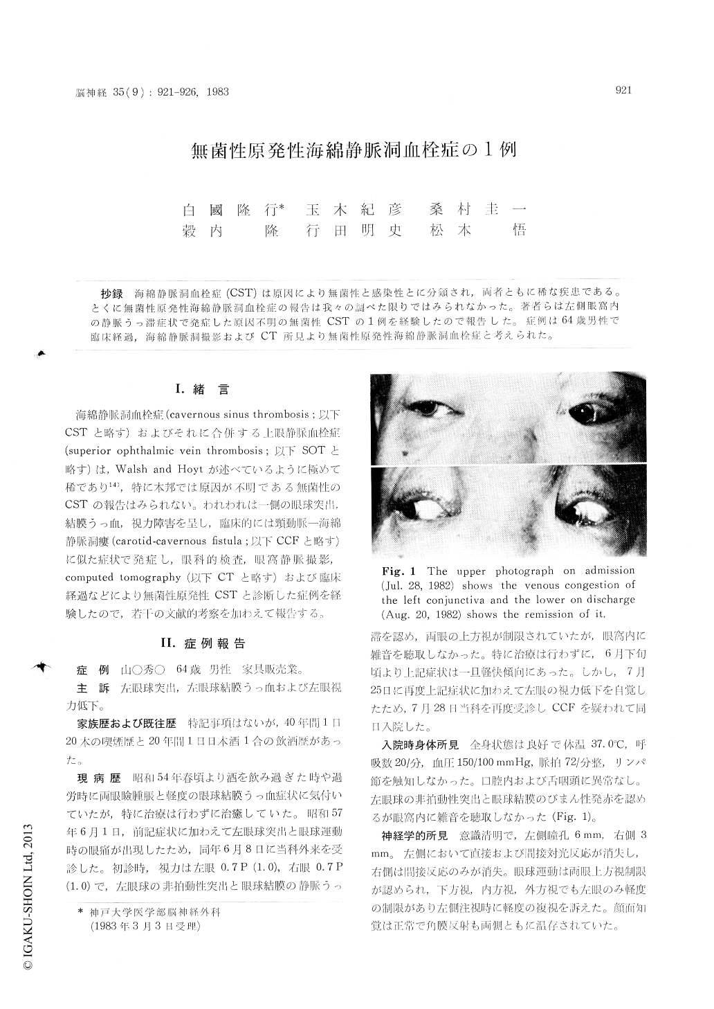
This 64 years male admitted to our clinic with the complaints of non-pulsatile exophthalmosis and conjunctival congestion of left eye. On admis-sion, he showed mild external ophthalmoplegia and clinical evidence of intraorbital congestion (choked disc, retinal vein thrombosis, retinal hemorrhage) on the left side. The blood examina-tion, including the thyroid studies, revealed no abnormal findings except for mild anemia and increased ESR. In carotid angiography, there was occlusion of Sylvian vein and cavernous sinus in the affected side. Orbital venography and retro-grade jugular venography demonstrated the occlu-sion of superior ophthalmic vein, cavernous sinus and inferior petreous sinus in left side. CT scan revealed parasellar enhanced area in the normal pattern. Enhanced orbital CT scan revealed the hypertrophy of left external occular muscles and optic nerve with a tomogram of the dilatated superior ophthalmic vein.
Aseptic primary CST was diagnosed on the basis of clinical course, cavernous sinography and CT findings.
Copyright © 1983, Igaku-Shoin Ltd. All rights reserved.