Japanese
English
- 有料閲覧
PPV購入案内
1,320円 (1,200円+税10%) こちらは、375ページから381ページの文献です。購入には医書.jp本会員登録が必要です。
会員登録完了後に改めて上記「購入サイトへ」を選択してください。
または「購入サイトへ」選択後に「購入ログイン」、「新規会員登録」の順に進んでください。
会員登録について詳しくはをご確認ください。
- Abstract 文献概要
- 1ページ目 Look Inside
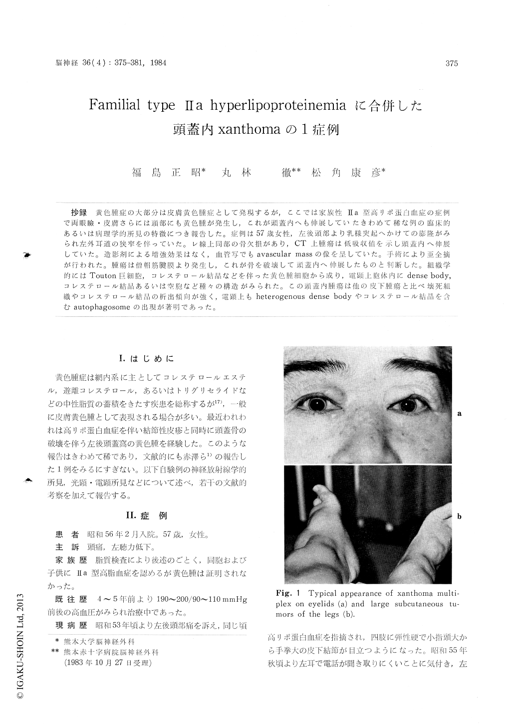
抄録 黄色腫症の大部分は皮膚黄色腫症として発現するが,ここでは家族性IIa型高リポ蛋白血症の症例で両眼瞼・皮膚さらには頭部にも黄色腫が発生し,これが頭蓋内へも伸展していたきわめて稀な例の臨床的あるいは病理学的所見の特徴につき報告した。症例は57歳女性,左後頭部より乳様突起へかけての膨隆がみられ左外耳道の狭窄を伴っていた。レ線上同部の骨欠損があり,CT上腫瘍は低吸収値を示し頭蓋内へ伸展していた。造影剤による増強効果はなく,血管写でもavascular massの像を呈していた。手術により亜全摘が行われた。腫瘍は僧帽筋腱膜より発生し,これが骨を破壊して頭蓋内へ伸展したものと判断した。組織学的にはTouton巨細胞,コレステロール結晶などを伴った黄色腫細胞から成り,電顕上胞体内にdense body,コレステロール結晶あるいは空胞など種々の構造がみられた。この頭蓋内腫瘍は他の皮下腫瘍と比べ壊死組織やコレステロール結晶の析出傾向が強く,電顕上もheterogenous dense bodyやコレステロール結晶を含むautophagosomeの出現が著明であった。
Familiar hyperlipoproteinemia associated with intracranial xanthoma belonged to a very rare entity and the authors were able to collect only one reported case in our domestic literature.
A 57-year-old house wife was admitted with a six months' history of progressive hearing impair-ment on the left and some cerebellar signs cha-racterized by ataxic gait. Physical examination revealed several hard and painful subcutaneous masses around the joints of four extremities, which had enlarged slowly within the last three years. Also, remarkable deformity of Achilles tendon was seen on both sides. Neurological examination showed bilateral papilledema, left hemifacial hyp-and dysesthesia, left hearing difficulty and left cerebellar signs.
Laboratory examination reported markedly ele-vated serum cholesterol level as high as 575mg/dl, and determination of the serum lipoprotein dis-closed the findings compatible with Type Ira hyperlipoproteinemia. Plain skull X-rays showed osteolytic defect of the left occipital bone and CT scan demonstrated a large irregular low density mass extending into the left posterior fossa, which showed ring like enhancement and spotty high density of calcification. Angiogram suggested the mass was of less vascular lesion and situated extramedullary. Suboccipital craniectomy was per-formed and an epidural solid mass was resected, although the mass was markedly growing into the posterior cranial cavity.
Microscopic examination showed the findings typical to the xanthoma, which was totally coin-cided with that of biopsy specimen obtained from the tumor over the ankle. Electron microscopic examination was performed and various kinds of lipid inclusions in the cytoplasma of xanthoma cells, such as dense bodies, volute or onion-like figures and lucent large vacuoles, were observed.
Copyright © 1984, Igaku-Shoin Ltd. All rights reserved.