Japanese
English
- 有料閲覧
PPV購入案内
1,320円 (1,200円+税10%) こちらは、437ページから442ページの文献です。購入には医書.jp本会員登録が必要です。
会員登録完了後に改めて上記「購入サイトへ」を選択してください。
または「購入サイトへ」選択後に「購入ログイン」、「新規会員登録」の順に進んでください。
会員登録について詳しくはをご確認ください。
- Abstract 文献概要
- 1ページ目 Look Inside
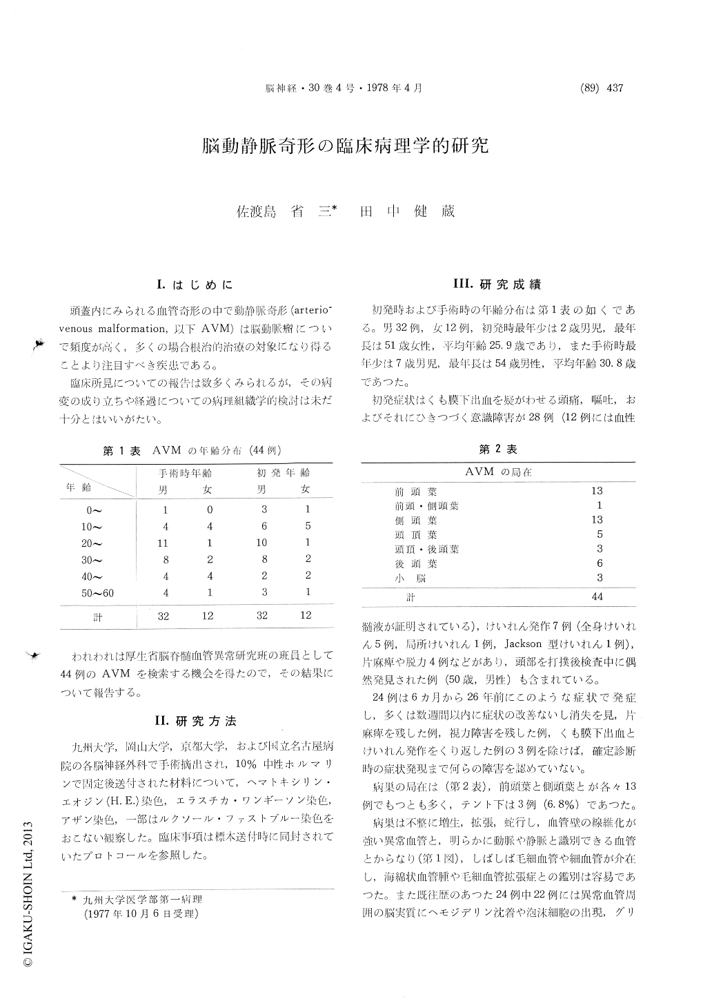
I.はじめに
頭蓋内にみられる血管奇形の中で動静脈奇形(arterio—venous malforrnation,以下AVM)は脳動脈瘤についで頻度が高く,多くの場合根治的治療の対象になり得ることより注目すべき疾患である。
臨床所見についての報告は数多くみられるが,その病変の成り立ちや経過についての病理組織学的検討は未だ十分とはいいがたい。
Forty-four cases of cerebral arteriovenous malfor-mation (AVM), 32 males and 12 females, were ex-amined clinicopathologically.
The average age is 25.9 years at the onset of symptoms and is 30.8 years at the time of surgical excision. There were 41 cases of supratentorial AVM and 3 were found below the tentorium, and 13 cases were found in the frontal and temporal areas respectively. Twenty-four among 44 cases suffered from cerebrovascular accidents or convulsive seizures 6 months to 20years before the final diagnosis. Among them 22 cases showed hemo-siderin deposition, foam cell accumulation and proliferation of glial cells and capillaries in the lesion of AVM, suggesting old hemorrhage or cerebral softening.
AVM consists of many dilated vessels of variable diameter. Fistulous communication of abnormal artery and vein is one of the most characteristic findings. More severe structural alterations of vessles such as dilatation or narrowing of the lumina, tortuosity and irregular fibrous thickening of the wall and duplication or loss of internal elastic lamina are present in veins than in arteries. Perivascular fibrosis is sometimes a feature. Mural thrombi showing organization or recanalization are detected in 5 cases. In the intervening or adjoyning brain parenchyma, there are often areas of con-taining many capillaries or small vessels which are considered to play an important part in pro-gression of AVM with advance of age. The rupture of the vessel wall is noted in 3 cases all of which are veins or venous abnormal vessels. Fibrinoid necrosis of the vessel wall is not evident.
Copyright © 1978, Igaku-Shoin Ltd. All rights reserved.