Japanese
English
- 有料閲覧
PPV購入案内
1,320円 (1,200円+税10%) こちらは、1343ページから1349ページの文献です。購入には医書.jp本会員登録が必要です。
会員登録完了後に改めて上記「購入サイトへ」を選択してください。
または「購入サイトへ」選択後に「購入ログイン」、「新規会員登録」の順に進んでください。
会員登録について詳しくはをご確認ください。
- Abstract 文献概要
- 1ページ目 Look Inside
I.はじめに
筋紡錘における神経終末の研究は盛んに行われており,それらの殆んどは銀染色による光顕的観察あるいは電顕的観察によっている。Cholinesterase (ChE)染色による筋紡錘神経終末の証明は,1956年Cöers3)らにより初めて猫で成された。以来20年間に,ヒトの骨格筋筋紡鍾については未だその記載がみられない。今回,著者は正常ヒト骨格筋筋紡鍾を用い,ChE染色により3種類のfusimotor ending (筋紡鍾運動神経終末)を分類することができた。また,NADH-Tetrazolium Reductase (NADH-TR)染色によるmitochondrial oxidative enzyme activityを用い,2種類のfusisensory ending (筋紡錘知覚神経終末)を分類することができたので報告する。
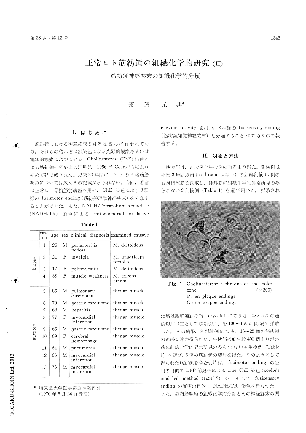
Muscle specimens were obtained from nine fresh autopsy cases (64-86 y. o.) and four biopsy cases (17-38 y. o.). The thenar muscles (autopsy) and other skeletal muscles (biopsy) were used (Table 1). These muscles had never histochemically patho-logical findings in the extrafusal muscles. Muscle tissues were quickly frozen in isopentane at -60 ℃ and cut into 10-15μ thick serial transverse sections in a cryostat, and then mounted on cover-slips or slides.
For histochemical demonstration of the intrafusal motor nerve endings, cholinesterase technique (Koelle's modified method) was employed and follow-ing three types of the fusimotor endings were classified (Table 3a & Fig. 6).
1) En plaque endings were observed as a plate-shaped cholinesterase activity on transverse sections(Fig. 1). This type of the fusimotor endings were localized at the polar zone on Bag II fibers and Chain II fibers, which are histochemical types of the intrafusal muscle fibers and were described previously by auther.
2) En grappe endings were seen as an arched cholinesterase activity on transverse sections (Fig. 1). This type of the fusimotor endings were localized at the polar zone on both the nuclear bag fiber (nbf) and the nuclear chain fiber (ncf).
3) Diffuse endings were observed as a ring-shaped cholinesterase activity on transverse sections (Fig. 2). This type of the fusimotor endings were localized at the intermediate zone on both nbf and ncf.
For histochemical demonstration of the fusisensory endings, cholinesterase technique were employed, but negative cholinesterase activity were revealed at the equatorial zone (Fig. 3). So using mitochondrial oxidative enzyme activity of NADH-TR stain, following two types of the fusisensory endings were classified (Table 3a & Fig, 6).
1) Primary sensory endings were observed as large irregular patches of diformazan. This type of the fusisensory endings were localized at the juxta-equatorial zone on both nbf and ncf (Fig. 4a). And then, this large irregular patches of diformazan well corresponded electron-microscopically to aboundant mitochondrial distribution of the primary sensory ending (Fig. 4b).
2) Secondary sensory endings were seen as a halo-shaped enzyme activity of diformazan (Fig. 5a). This type of the fusisensory endings were localized at the equatorial zone on both nbf and ncf. And then, this halo of diformazan well corresponded electronmicroscopically to scarce mitochondrial dis-tribution of the secondary sensory ending (Fig. 5b).
Copyright © 1976, Igaku-Shoin Ltd. All rights reserved.