Japanese
English
- 有料閲覧
- Abstract 文献概要
- 1ページ目 Look Inside
I.はじめに
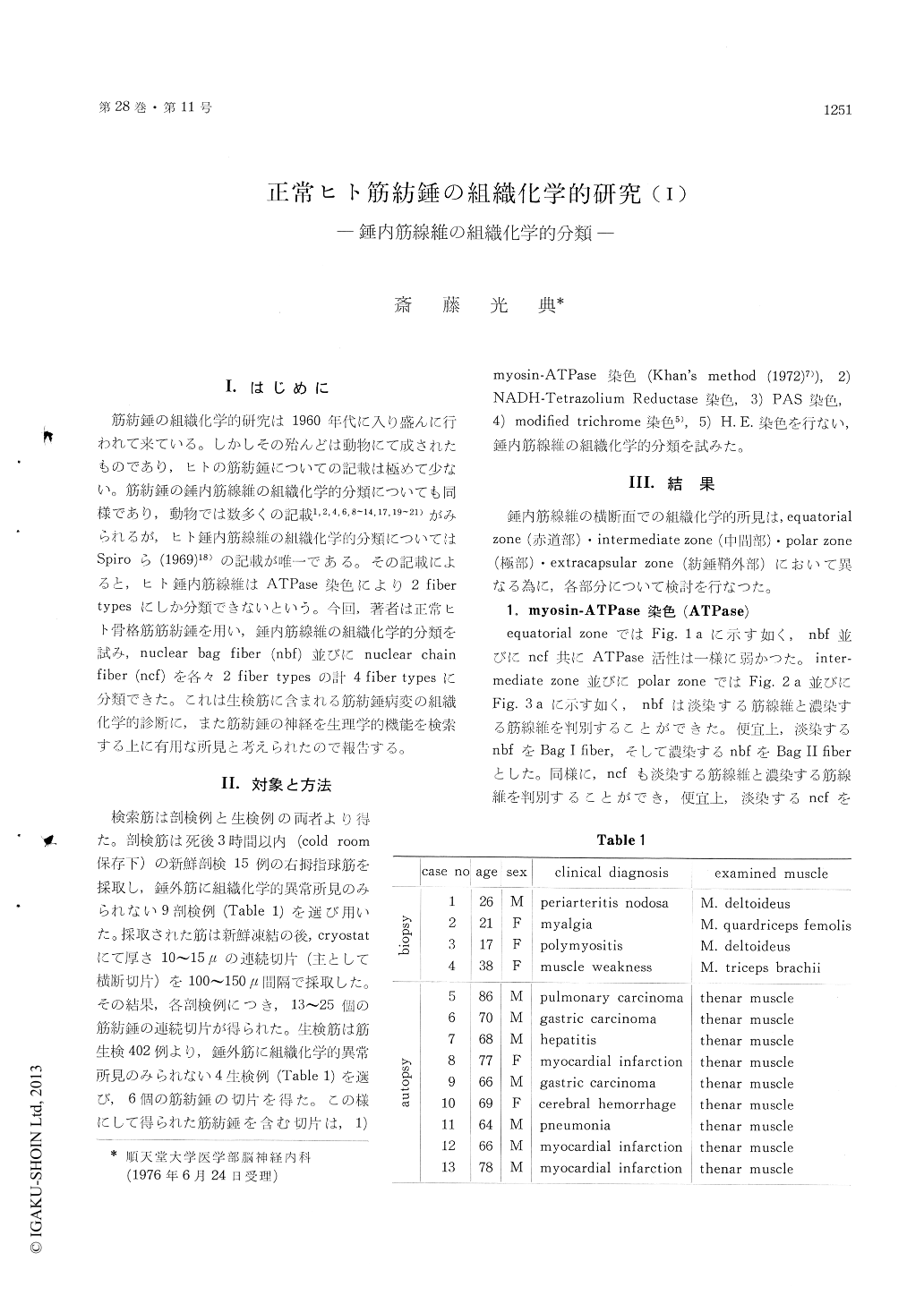
筋紡錘の組織化学的研究は1960年代に入り盛んに行われて来ている。しかしその殆んどは動物にて成されたものであり,ヒトの筋紡錘についての記載は極めて少ない。筋紡錘の錘内筋線維の組織化学的分類についても同様であり,動物では数多くの記載1,2,4,6,8〜14,17,19〜21)がみられるが,ヒト錘内筋線維の組織化学的分類についてはSpiroら(1969)18)の記載が唯一である。その記載によると,ヒト錘内筋線維はATPase染色により2 fibertypesにしか分類できないという。今同,著者は正常ヒト骨格筋筋紡錘を用い,錘内筋線維の組織化学的分類を試み,nuclear bag fiber(nbf)並びにnuclear chainfiber(ncf)を各々2 fiber typesの計4 fiber typesに分類できた。これは生検筋に含まれる筋紡錘病変の組織化学的診断に,また筋紡錘の神経を生理学的機能を検索する上に有用な所見と考えられたので報告する。
Muscle specimens were obtained from nine fresh autopsy cases (64-86 y. o.) and four biopsy cases (17-38 y. o.). The thenar muscles (autopsy) and other skeletal muscles (biopsy) were used (Table 1). These muscles had never histochemically patho-logical findings in the extrafusal muscles. Muscle tissues were quickly frozen in isopentane at -60℃ and cut into 10-15 thick serial transverse sections in a cryostat, and then mounted on coverslips or slides. For histochemical demonstration of the intra-fusal muscle fibers, myosin-ATPase stain (Khan's method), NADH-Tetrazolium Reductase stain, PAS stain, modified trichrome stain and H. E. stain were employed.
As the result of histochemical study, the four types of human intrafusal muscle fibers were classi-fied. Using myosin-ATPase reaction, four types of the intrafusal muscle fibers were noted at the intermediate and polar zone (Table 2b & Fig. 7a). In these zone, nuclear bag fibers (nbf) were classi-fied two kinds of ATPase-light-stained fiber and ATPase-dark-stained fiber (Fig. 2a & Fig. 3a), tentatively ATPase-light-stained bag fiber was named as "Bag I fiber" and ATPase-dark-stained bag fiber was named as "Bag II fiber". Nuclear chain fibers (ncf) were similarly classified two kinds of ATPase-light-stained fiber and ATPase-dark-stained fiber (Fig. 2 a & Fig. 3 a), tentatively ATPase-light-stained chain fiber was named as "Chain I fiber" and ATPase-dark-stained chain fiber was named as "Chain II fiber". This "Chain I fiber" was noted rarely.
By the mitochondrial oxidative enzymatic activity of NADH-TR stain, "Bag I fiber" showed dark stain with fine diformazan granulation and "Bag II fiber" showed light stain with rough diformazan granulation (Fig. 7 b), and then, ncf revealed uniformly wheel-like distribution of diformazan granulation (Fig. 2 b & Fig. 3 b).
With PAS reaction, "Bag I fiber" showed fine glycogen granulation and "Bag II fiber" showed rough glycogen granulation, and then, ncf revealed uniformly wheel-like distribution of glycogen granulation (Fig. 5).

Copyright © 1976, Igaku-Shoin Ltd. All rights reserved.


