Japanese
English
- 有料閲覧
PPV購入案内
1,320円 (1,200円+税10%) こちらは、67ページから76ページの文献です。購入には医書.jp本会員登録が必要です。
会員登録完了後に改めて上記「購入サイトへ」を選択してください。
または「購入サイトへ」選択後に「購入ログイン」、「新規会員登録」の順に進んでください。
会員登録について詳しくはをご確認ください。
- Abstract 文献概要
- 1ページ目 Look Inside
I.はじめに
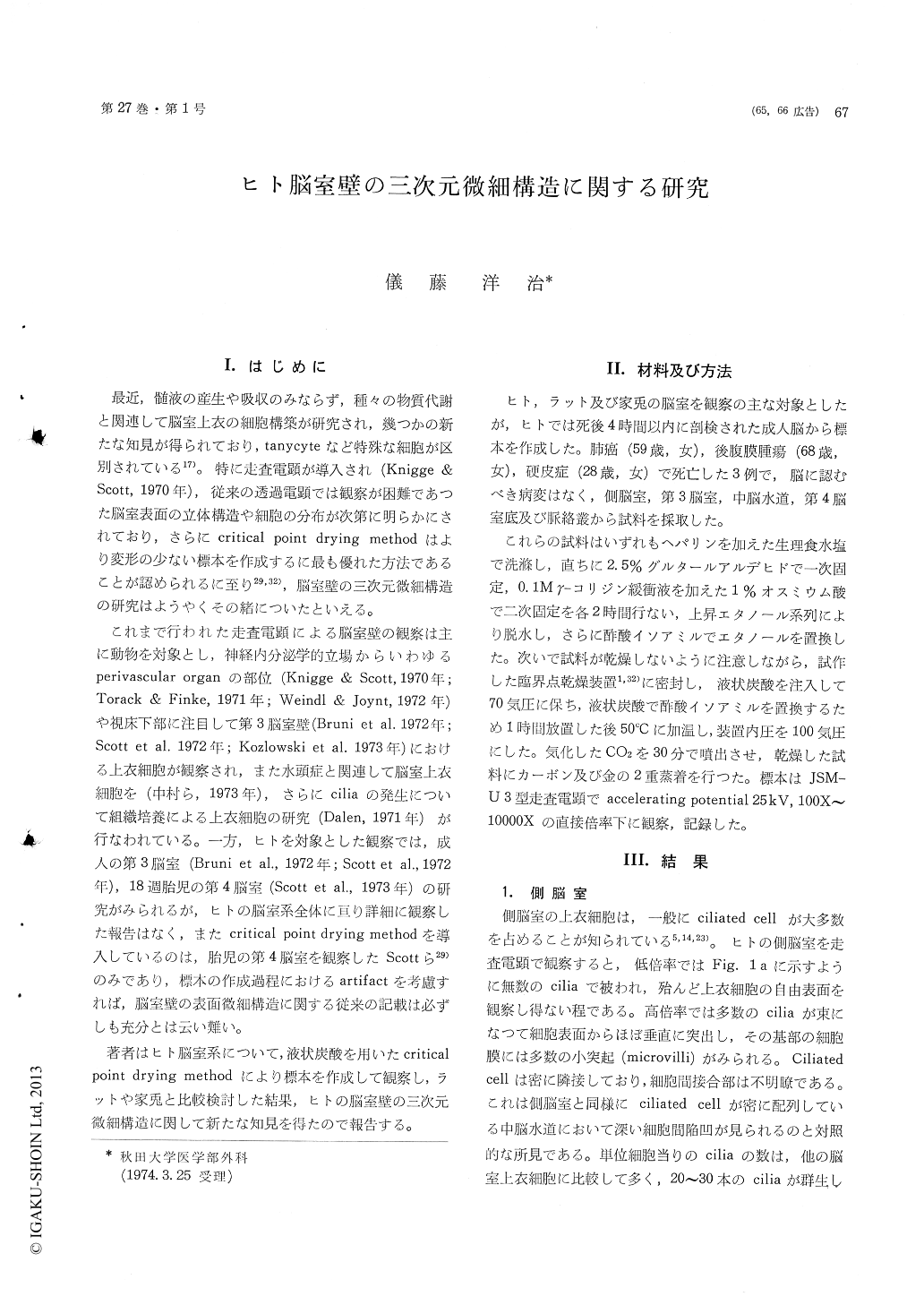
最近,髄液の産生や吸収のみならず,種々の物質代謝と関連して脳室上衣の細胞構築が研究され,幾つかの新たな知見が得られており,tanycyteなど特殊な細胞が区別されている17)。特に走査電顕が導入され(Knigge &Scott,1970年),従来の透過電顕では観察が困難であつた脳室表面の立体構造や細胞の分布が次第に明らかにされており,さらにcritical point drying methodはより変形の少ない標本を作成するに最も優れた方法であることが認められるに至り29,32),脳室壁の三次元微細構造の研究はようやくその緒についたといえる。
これまで行われた走査電顕による脳室壁の観察は主に動物を対象とし,神経内分泌学的立場からいわゆるperivascular organの部位(Knigge & Scott,1970年;Torack & Finke,1971年;Weindl & Joynt,1972年)や視床下部に注目して第3脳室壁(Bruni et al.1972年;Scott et al.1972年;Kozlowski et al.1973年)における上衣細胞が観察され,また水頭症と関連して脳室上衣細胞を(中村ら,1973年),さらにciliaの発生について組織培養による上衣細胞の研究(Dalen,1971年)が行なわれている。一方,ヒトを対象とした観察では,成入の第3脳室(Bruni et al.,1972年;Scott et al.,1972年),18週胎児の第4脳室(Scott et al.,1973年)の研究がみられるが,ヒトの脳室系全体に亘り詳細に観察した報告はなく,またcritical point drying methodを導入しているのは,胎児の第4脳室を観察したScottら29)のみであり,標本の作成過程におけるartifactを考慮すれば,脳室壁の表面微細構造に関する従来の記載は必ずしも充分とは云い難い。
The regional structural differences of the humancerebral ventricle have been investigated with thescanning electron microscopy from the point offunctional specialization in the ventricular system.
Materials have been exercised, beginning withthe normal adult human brain at autopsy. Blocksof tissue have been dehydrated in graded alcoholsand amyl-acetate after double fixation in 2. 5%glutar-aldehyde and in 1% 0s04 solution added0.1 Mr-collidin buffer. Tissues have been proceededby a critical point drying method and coated withcarbon and gold in a vaccum evaporater. Thesurface of specimens has been examined with JSM-U3 scanning electron microscope. The results ofobservation are as follows. Ependymal cilia of thehuman brain extend in clusters into the ventricleat the apical surfaces of the ependymal cells.These have an average diameter of 0.25μ andlength of 10μ. Human cilia taper towards thetip like those of rabbit, while club-form in rat.
The lateral ventricle and the cerebral aqueductare so much covered with numerous ciliated epen-dymal cells that the observation of plasmalemmais rather difficult. In the cerebral aqueduct, inter-ependymal gaps are deep, in contrast to the lateralventricle where there are superficial intercellularfurrows.
The human third ventricle exhibits the regionalvariation in the surface structure as seen in animals.Ciliated ependymal cells become sparce towardsthe infundibular recess. The transition zone occursat the level of the underlying paraventicular nuc-leus in the human brain, while it occurs at thelevel of dorsomedial nucleus in rat and rabbit.
A number of hemispherical cells, devoiding ciliaand microvilli, are observed among ciliated epen-dymal cells near the apertura lateralis in the humanfourth ventricle.
The human choroid plexus shows a typical con-voluted structure. Most of epithelial cells possessslender and short cilia. A profusion of microvilliare observed on the surface of the epithelial cells.Most numerous microvilli are observed at thechoroid plexus in the human brain.
Copyright © 1975, Igaku-Shoin Ltd. All rights reserved.