Japanese
English
- 有料閲覧
- Abstract 文献概要
- 1ページ目 Look Inside
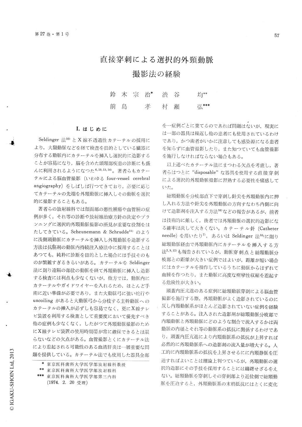
I.はじめに
Seldinger法10)とX線不透過性カテーテルの採用により,大腿動脈などを経て検査を目的としている臓器に分布する動脈内にカテーテルを挿入し選択的に造影することが容易になり,脳を含めた頭頸部疾患の診断にも盛んに利用されるようになつた6,10,13,14)。著者らもカテーテルによる脳血管撮影(いわゆるfour-vessel cerebralangiography)をしばしば行つてきており,必要に応じてカテーテルの先端を外頸動脈に挿入しその動脈を選択的に撮影することもある。
著者らの放射線科では顔面部の悪性腫瘍や血管腫の症例が多く,それ等の診断や放射線治療方針の決定やプランニングに選択的外頸動脈撮影の所見が重要な役割をはたしてきている。Scheunemann&Schrudde11)のように浅側頭動脈にカテーテルを挿入し外頸動脈を造影する方法は抗癌剤の動脈内持続注入療法中に採用することはあつても,純粋に診断を目的とした場合には手技そのものが繁雑すぎるきらいがある。カテーテルをSeldinger法に則り遠隔の部位の動脈を経て外頸動脈に挿入し造影する検査には利点も少なくないが,他方では,動脈内にカテーテルやガイドワイヤーを入れるため,ほとんど手術に近い準備が必要であり,また大動脈弓に強い蛇行やuncoilingがあると大動脈弓から分枝する主幹動脈へのカテーテルの挿入が必ずしも容易でなく,更にX線テレビ装置を利用する検査として重要度において優先すべき他の症例も少なくなく,したがつて外頸動脈撮影のためにX線テレビ装置の使用時間帯が常に確保できるとは限らないなどの欠点がある。血管撮影とくにカテーテル法により惹起される可能性のある血清肝炎は一層重要な問題を提供している。カテーテル法でも使用した器具全部を一症例ごとに棄てるのであれば問題はないが,現実には一部の器具は繰返し他の患者にも使用されているわけであり,かつ術者がいかに注意しても感染源になる患者を知らずに血管撮影したり,また知つていても血管撮影を施行しなければならない場合もある。
The authors have attempted thirty-five externalcarotid angiographies in the direct puncture methodin a consecutive series of thirty-three cases of thevarious diseases of the head, including three casesof meningioma. The attempt was successful inthirty angiographies; the rate of success was 86per cent.
In two of the five failures, the angiographersatisfied himself to do the common carotid angio-graphy, after the first trial resulted in the internalcarotid angiography. In the third and fourth fail-ures, the angiographer could not help to insertthe needle into the uppermost portion of thecommon carotid artery, since the malignant meta-stases to the submandibular lymph-nodes preventedhim from selecting the proper site of needling. Inthe last failure, the attempt of the external carotidangiography was given up because of the patient'sincooperation.
The authors are now convinced that the selec-tive external carotid angiography in the directpuncture method might be performed in as goodrate of success as the Ruggiero's one, if the inade-quate cases for the angiography as stated abovewould be excluded.
Three cases of meningioma, of which roentgeno-graphic confirmation was made based upon theselective external carotid angiogram in the directpuncturing method, are presented. The middlemeningeal artery, the feeding artery to the tumor,was markedly, dilated in caliber and markedlytortuous in course, and the characteristic patternof vascularity in meningioma was obtained in allthree cases. In the first case, a huge, extremelyhypervascularized tumor had been found in thefrontal region of the left cerebral hemisphere onthe internal carotid angiogram, carried out atanother institute, and the external carotid angio-graphy was attempted by the authors in order todifferentiate between glioblastoma and meningioma.In the remaining two cases, the external carotidangiography was done to confirm that the tumorwas supplied blood mainly from the middle menin-geal artery.
Since the external carotid angiography in thedirect puncturing method is not so difficult as be-ing stressed by many authors but Ruggiero andthe angiography in the catheterization method maynot always be possible to be performed because ofvarious reasons, the angiographer dealing with thediseases of the head and face should master theexternal carotid angiography in the direct punc-turing method.

Copyright © 1975, Igaku-Shoin Ltd. All rights reserved.


