Japanese
English
- 有料閲覧
PPV購入案内
1,320円 (1,200円+税10%) こちらは、595ページから600ページの文献です。購入には医書.jp本会員登録が必要です。
会員登録完了後に改めて上記「購入サイトへ」を選択してください。
または「購入サイトへ」選択後に「購入ログイン」、「新規会員登録」の順に進んでください。
会員登録について詳しくはをご確認ください。
- Abstract 文献概要
- 1ページ目 Look Inside
はじめに
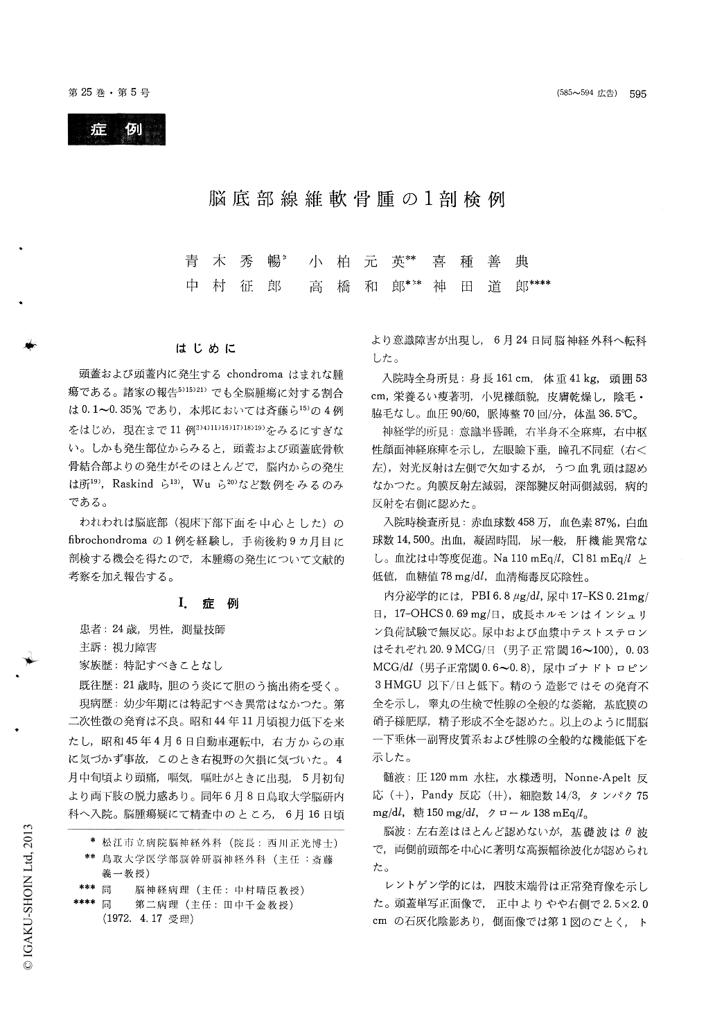
頭蓋および頭蓋内に発生するchondromaはまれな腫瘍である。諸家の報告5)15)21)でも全脳腫瘍に対する割合は0.1〜0.35%であり,本邦においては斉藤ら15)の4例をはじめ,現在まで11例3)4)11)16)17)18)19)をみるにすぎない。しかも発生部位からみると,頭蓋および頭蓋底骨軟骨結合部よりの発生がそのほとんどで,脳内からの発生は所19), Raskindら13),Wuら20)など数例をみるのみである。
われわれは脳底部(視床下部下面を中心とした)のfibrochondromaの1例を経験し,手術後約9カ月目に剖検する機会を得たので,本腫瘍の発生について文献的考察を加え報告する。
1) Intracranial and calvarial chondromas have been rarely reported. They usually have appeared to arise from the synchondrosis of the base of the skull.
A 24 year-old male was admitted to our clinic on June 24, 1970, because of disturbance of cons-ciousness. Physical examinations were as followed ; dehydration, semicomatose state, left hemiparesis with pathologic relexes, anisocoria (rt<lt), absent of left light reflex without chocked disc. The find-ings on endocrinological examinations revealed general dysfunction of diencephalo-hypophyseal system, adrenal and sexual glands.
Plain craniogram showed definite calcification inthe suprasellar region (2.5 × 2.0cm in size) with-out destruction of the anterior and posterior clinoid processes and clivus. Carotid and vertebral angio-graphies revealed a space-occupying lesion in the supra-and parasellar regions from the base of the skull.
A bifrontal craniotomy was performed on July 6, 1970. A hard nodular tumor was observed in the chiasmal region, extending posteriorly to the interpeduncular fossa. Partial removal of the tumor was followed by radiotherapy of 60Co.
2) At autopsy, a large tumor (5 × 4 × 5cm in size) was found in the base of the brain without involv-ing the dura and synchondrosis of the base of theskull. It was bony in consistencty and well de-marcated with circumscribed parts of brownish appearance. The bony portion of the tumor showed histologically feature of benign chondroma with myxoid degeneration in a part and the circums-cribed portions contained fibroma.
3) Cartilaginous tumors arising from the intra-cranial structures are of rare occurrence. It appears that our case originates from residual nests of embryonic cartilaginous tissue of the base of the brain. The origin and nature of this tumor were discussed.
Copyright © 1973, Igaku-Shoin Ltd. All rights reserved.