Japanese
English
- 有料閲覧
PPV購入案内
1,320円 (1,200円+税10%) こちらは、380ページから385ページの文献です。購入には医書.jp本会員登録が必要です。
会員登録完了後に改めて上記「購入サイトへ」を選択してください。
または「購入サイトへ」選択後に「購入ログイン」、「新規会員登録」の順に進んでください。
会員登録について詳しくはをご確認ください。
- Abstract 文献概要
- 1ページ目 Look Inside
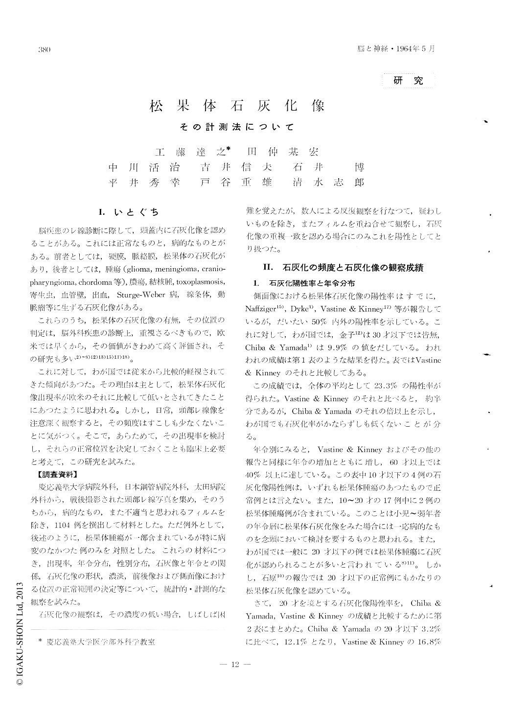
I.いとぐち
脳疾患のレ線診断に際して,頭蓋内に石灰化像を認めることがある。これには正常なものと,病的なものとがある。前者としては,硬膜,脈絡膜,松果体の石灰化があり,後者としては,腫瘍(glioma, meningioma, cranio—pharyngioma, chordoma等),膿瘍,結核腫,toxoplasmosis,寄生虫,血管壁,出血,Sturge-Weber病,線条体,動脈瘤等に生ずる石灰化像がある。
これらのうち,松果体の石灰化像の有無,その位置の判定は,脳外科疾患の診断上,重視さるべきもので,欧米では早くから,その価値がきわめて高く評価され,その研究も多い2)〜8)12)13)15)17)18)。
Normal skull X-ray skull films (1104 cases) are investigated and 23.3% of them show pineal body calcification. Several statistical studies are done (sex, age, size and concentration etc.).
New measuring method of pineal body calcification is devised in the lateral view of the skull X-ray film which is determined by the distances between the calcification and the center of sella turcica, bregma, internal occipital protuberance and the external acoustic meatus. Normal limits of the distances which mentioned as above are 4.0~5.0 cm, 6.5~8.0 cm, 7.5~9.0 cm, 6.0~7.5 cm and 3.5~ 5.0 cm.
Copyright © 1964, Igaku-Shoin Ltd. All rights reserved.