Japanese
English
- 有料閲覧
- Abstract 文献概要
- 1ページ目 Look Inside
はじめに
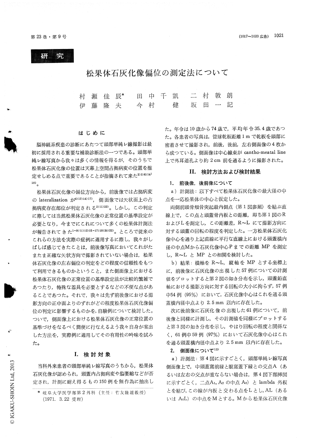
脳神経系疾患の診断にあたつて頭部単純レ線撮影は最初に採用される重要な補助診断法の一つである。頭部単純レ線写真から我々は多くの情報を得るが,そのうちで松果体石灰化像の位置は天幕上空間占拠病変の位置を推定せしめる点で重要であることが指摘されて来た2)3)6)14)18)。
松果体石灰化像の偏位方向から,前後像では占拠病変のlateralizationが4)12)14)17),側面像では矢状面上の占拠病変存在部位が判定される2)11)22)。しかし,この判定に際しては当然松果体石灰化像の正常位置の基準設定が必要となり,今までにこれについて多くの松果体計測法が報告されてきた1〜9)11)12)15〜17)19)20)22)。ところで従来のこれらの方法を実際の症例に適用するに際し,我々がしばしば感じてきたことは,前後像写真においてこれがたまたま正確な矢状方向で撮影されていない場合は,松果体石灰化像の左右偏位の判定をどの程度の信頼性をもつて利用できるものかということ,また側面像上における松果体石灰化像の正常位置の基準設定法が比較的繁雑であつたり,特殊な器具を必要とするなどの不便な点があることであつた。それで,我々は先ず前後像における撮影方向の正中面よりのずれがどの程度松果体石灰化像偏位の判定に影響するものかを,自験例について検討した。ついで,側面像上における松果体石灰化像の正常位置の基準づけをなるべく簡便に行なえるよう我々自身が案出した方法を,実際例に適用してその有用性の吟味を試みた。
Pineal calcification in plain craniogram is a useful landmark for estimating location of intracranial space-occupying lesions. Various methods of measur-ing its standard position hitherto have been reported. However, in applying these methods practically, inconveniences sometimes may be encountered. One of the inconveniences is that, when A-P or P-A film is made while the head happened to be some-what rotated around the vertical axis, lateral shift of pineal calcification may be difficult to be deter-mined. Another problem is that the known methods of measuring the position of pineal calcification in lateral film are relatively complicated or incon-venient. In this report we studied influence of slight head rotation on estimation of lateral shift of pineal calcification in A-P or P-A film, using craniograms of 150 normal subjects.
Study was further performed on a simple method of measuring pineal calcification in lateral film, which was devised by ourselves. The following results were obtained.
1) In normal subjects, the center of pineal cal-cification in A-P or P-A film existed within 2. 5 mm in either direction from the midpoint of the inner horizontal skull diameter passing through the pineal calcification, irrespective of slight rotation of the head around the vertical axis. Location of the calcification outside this range will be pathological.
2) In normal subjects, the center of pineal calci-fication in lateral film existed near the midpoint M of the line AL (or A0L). The line AL (or A0L) is defined as follows. The crossing point A of the curved line, indicating anterior border of the middle cranial fossa, and the line, indicating lower surface of the orbital roof, (or the midpoint A0 of the crossing point A1 and A2 on both sides) is connected by a line to outer lambda. and the point at which the inner table is crossed is named L. In most of the cases the center of pineal calcification was found to exist inside a square of 12 ×1.2 mm2, of which the center was located 4 mm anterior to and 2 mm below the point M. In cases where lambda was hardly identified, it was found that the point L could be substituted by the point L', which was a point on the inner table 10 cm distant from the most caudal point B on the inner table of the occipital bone. Location of the center of pineal calcification outside this square probably will be pathological.

Copyright © 1971, Igaku-Shoin Ltd. All rights reserved.


