Japanese
English
- 有料閲覧
PPV購入案内
1,320円 (1,200円+税10%) こちらは、1341ページから1345ページの文献です。購入には医書.jp本会員登録が必要です。
会員登録完了後に改めて上記「購入サイトへ」を選択してください。
または「購入サイトへ」選択後に「購入ログイン」、「新規会員登録」の順に進んでください。
会員登録について詳しくはをご確認ください。
- Abstract 文献概要
- 1ページ目 Look Inside
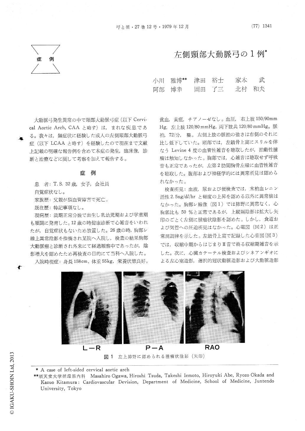
大動脈弓発生異常の中で頸部大動脈弓症(以下Cervi—cal Aortic Arch, CAAと略す)は,まれな疾患である。我々は,無症状に経験した成人の左側頸部大動脈弓症(以下LCAAと略す)を経験したので現在まで文献上記載の明確な報告例を含めて本症の発生,臨床像,診断と治療などに関して考察を加えて報告する。
A case of left-sided cervical aortic arch was reported. A 32-year-old female, asymptomatic, was admitted to our hospital for an evaluation of abnormality in chest X-ray film in January, 1979. In her childhood, she was noticed to have abnormal shadow in chest X-ray films, but remained untreated because of asymptomatic. On admission, the physical examination revealed that the bloo dpressure at right upper extremity was elevated more than that of left side by 30 mmHg. At left supraclavicular fossa, grade 4 out of 6 vascular murmur was heard with thrill. Pulsating mass, however, was not noticed there. Although a homogeneous mass shadow was seen in left upper lung field in chest X-ray film, there were no evidence of compression to neighbor organs. The cardiac catheterization data was unremarkable. The aortography demon-strated that there was slight dilatation of ascending aorta with right brachiocephalic trunk as the first branch from aortic arch, and left subclavian artery with it's root stenosis as the last one. The top of the arch was displaced toward left side of neck. These findings were identical with left-sided cervical aortic arch without associated cardiac anomaly. The cervical aortic arch is very rare congenital malformation. According to the literature, there have been 14 reported cases excluding the present case. The cases older than 30 years of age at the diagnosis are 4 among them, The clinical manifestations of dyspnea or dysphagia are derived from com-pressed or deviated trachea or esophagus by cervical aortic arch.
Copyright © 1979, Igaku-Shoin Ltd. All rights reserved.