Japanese
English
- 有料閲覧
PPV購入案内
1,320円 (1,200円+税10%) こちらは、1273ページから1279ページの文献です。購入には医書.jp本会員登録が必要です。
会員登録完了後に改めて上記「購入サイトへ」を選択してください。
または「購入サイトへ」選択後に「購入ログイン」、「新規会員登録」の順に進んでください。
会員登録について詳しくはをご確認ください。
- Abstract 文献概要
- 1ページ目 Look Inside
- 参考文献 Reference
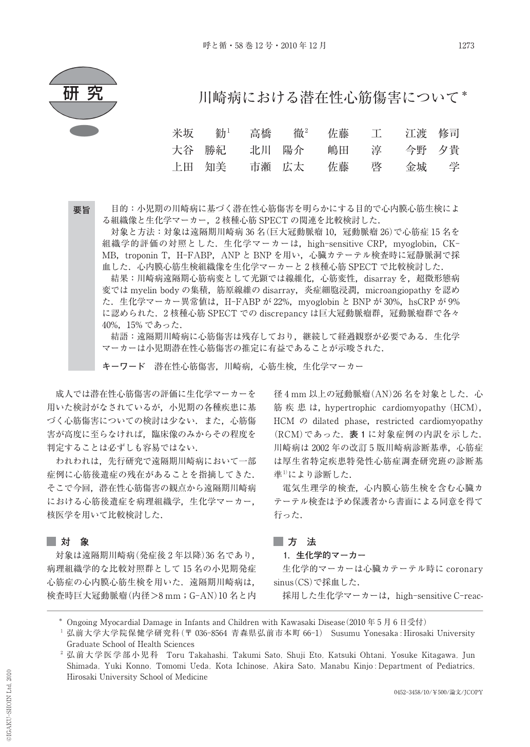
要旨 目的:小児期の川崎病に基づく潜在性心筋傷害を明らかにする目的で心内膜心筋生検による組織像と生化学マーカー,2核種心筋SPECTの関連を比較検討した.
対象と方法:対象は遠隔期川崎病36名(巨大冠動脈瘤10,冠動脈瘤26)で心筋症15名を組織学的評価の対照とした.生化学マーカーは,high-sensitive CRP,myoglobin,CK-MB,troponin T,H-FABP,ANPとBNPを用い,心臓カテーテル検査時に冠静脈洞で採血した.心内膜心筋生検組織像を生化学マーカーと2核種心筋SPECTで比較検討した.
結果:川崎病遠隔期心筋病変として光顕では線維化,心筋変性,disarrayを,超微形態病変ではmyelin bodyの集積,筋原線維のdisarray,炎症細胞浸潤,microangiopathyを認めた.生化学マーカー異常値は,H-FABPが22%,myoglobinとBNPが30%,hsCRPが9%に認められた.2核種心筋SPECTでのdiscrepancyは巨大冠動脈瘤群,冠動脈瘤群で各々40%,15%であった.
結語:遠隔期川崎病に心筋傷害は残存しており,継続して経過観察が必要である.生化学マーカーは小児期潜在性心筋傷害の推定に有益であることが示唆された.
Background:Ongoing myocardial damage in infants and children has not been well defined. Furthermore, it is not so easy to estimate the severity of myocardial damage only from clinical symptoms and signs. To clarify the ongoing myocardial damage, biochemical markers, dual isotope myocardial single photon emission computed tomography(SPECT)were compared with endomyocardial biopsy(EMB)in patients with late stage Kawasaki disease(KD).
Patients and method:They included 36KD cases with coronary arterial lesions(CAL)including 10 of giant coronary aneurysms and 26 of coronary aneurysms, aged from 2.5 to 18 years, and 15 patients with cardiomyopathy(CM), aged from 3 to 15 years. Selected biochemical markers in this study were high-sensitive C-reactive protein(hsCRP), myoglobin, Creatin Kinase MB(CK-MB), troponin T(TnT), heart-type fatty acid binding protein(H-FABP), ANP and BNP. These biochemical markers were obtained from the coronary sinus during cardiac catheterization. Histopathology was evaluated using the histomorphometric method. The fraction of myocardial volume occupied by fibrillar collagen(the percent area of fibrosis;%fibrosis)and diameter of myocytes were detected by quantitative morphometry with an automated image analysis system in sections stained with AZAN stain. Histopathological assessment was made according to the diagnostic guidelines of the Pathological Branch of the Idiopathic Cardiomyopathy Research Committee of the Ministry of Health and Welfare of the Japanese Government. A scale of 0 to 3+ was used with 0 considered normal, 1+ mildly abnormal, 2+ moderately abnormal, and 3+ severely abnormal. In this study we regarded more than 1+ as significant. Discrepancy was diagnosed when mismatch was found in dual SPECT.
Results:Myocardial changes on EMB showed various histopathological findings such as degeneration, interstitial fibrosis and disarray. As ultrastructural changes, accumulation of myelin bodies, disarray of myofibrils, inflammatory cell infiltration and microangiopathy were shown. Prevalence of abnormal biochemical markers was found in 22% of H-FABP, 30% of myoglobin and BNP and 9% of hsCRP. Discrepancy of dual SPECT was shown in 40% of KD patients with giant coronary aneurysms and 18% of those with coronary aneurysms.
Conclusions:Discrepancy in dual SPECT images suggested that residual myocardial damage in long-standing KD with CAL might reflect not only myocardial ischemia but also disordered myocardial fatty acid metabolism following myocarditis. Selected biochemical markers may be available for predicting ongoing cardiomyocyte damage in the late stage of KD.
Copyright © 2010, Igaku-Shoin Ltd. All rights reserved.