Japanese
English
- 有料閲覧
PPV購入案内
1,320円 (1,200円+税10%) こちらは、40ページから51ページの文献です。購入には医書.jp本会員登録が必要です。
会員登録完了後に改めて上記「購入サイトへ」を選択してください。
または「購入サイトへ」選択後に「購入ログイン」、「新規会員登録」の順に進んでください。
会員登録について詳しくはをご確認ください。
- Abstract 文献概要
- 1ページ目 Look Inside
- 参考文献 Reference
- サイト内被引用 Cited by
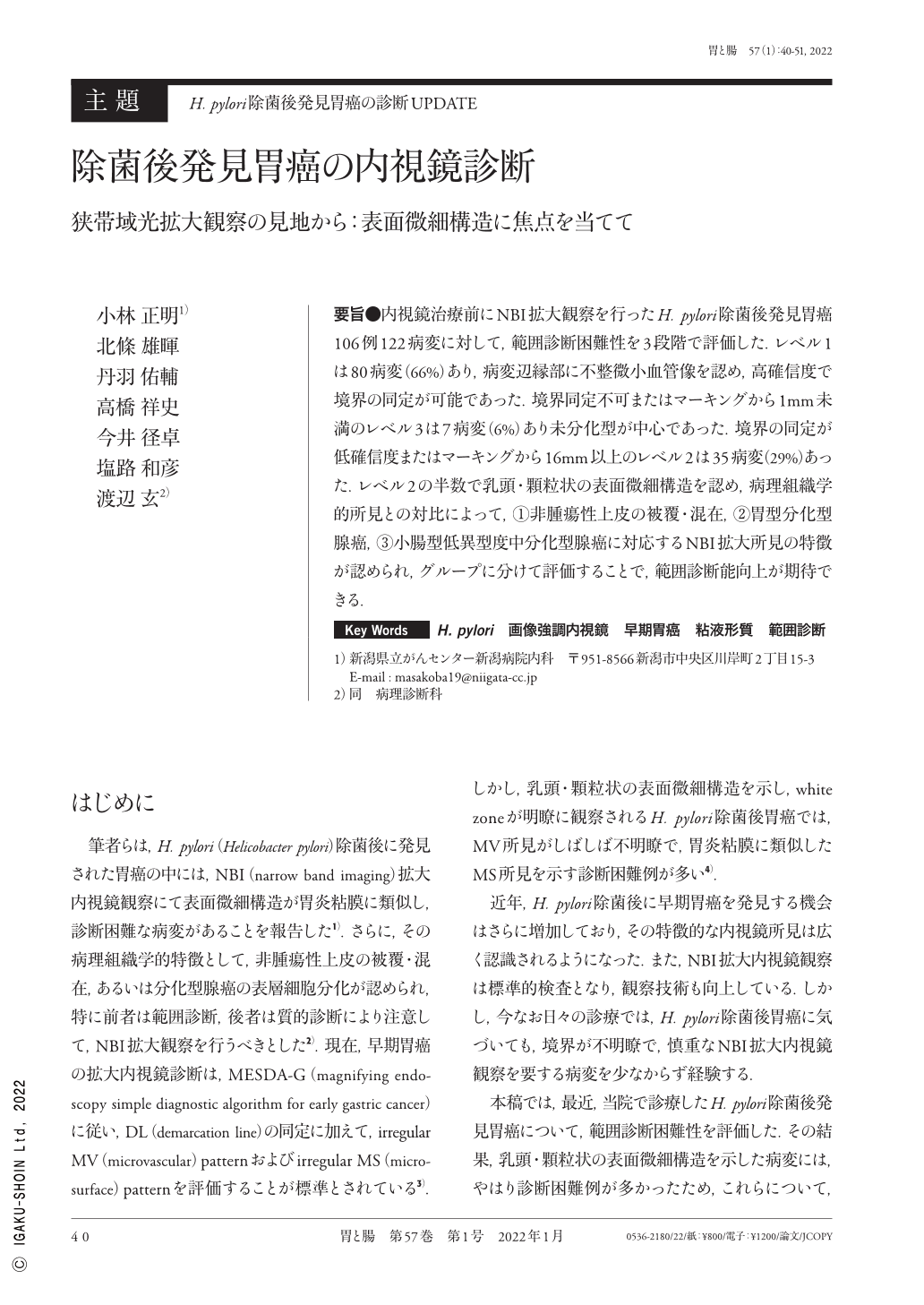
要旨●内視鏡治療前にNBI拡大観察を行ったH. pylori除菌後発見胃癌106例122病変に対して,範囲診断困難性を3段階で評価した.レベル1は80病変(66%)あり,病変辺縁部に不整微小血管像を認め,高確信度で境界の同定が可能であった.境界同定不可またはマーキングから1mm未満のレベル3は7病変(6%)あり未分化型が中心であった.境界の同定が低確信度またはマーキングから16mm以上のレベル2は35病変(29%)あった.レベル2の半数で乳頭・顆粒状の表面微細構造を認め,病理組織学的所見との対比によって,①非腫瘍性上皮の被覆・混在,②胃型分化型腺癌,③小腸型低異型度中分化型腺癌に対応するNBI拡大所見の特徴が認められ,グループに分けて評価することで,範囲診断能向上が期待できる.
We evaluated the microvascular and microsurface pattern of 122 early gastric cancers detected in 106 patients who received successful Helicobacter pylori eradication therapy. The diagnostic difficulty of demarcation line using NBI-ME(magnifying endoscopy with narrow band imaging)for gastric cancers after eradication was ranked in three levels. High confidence(level 1, n=80)was obtained in cancers with an irregular microvascular pattern. Low confidence(level 3, n=7)was mainly observed in undifferentiated cancers. The papillary microstructure that resembled the adjacent noncancerous mucosa was observed in approximately 50% of the cancers with middle confidence(level 2, n=35). NBI-ME revealed the following pathological findings in cancers with papillary microstructures ; (1)non-neoplastic superficial epithelium and(2)surface differentiation of tubular adenocarcinoma with gastric or small intestinal mucin phenotypes.
Copyright © 2022, Igaku-Shoin Ltd. All rights reserved.