Japanese
English
- 有料閲覧
PPV購入案内
1,320円 (1,200円+税10%) こちらは、96ページから104ページの文献です。購入には医書.jp本会員登録が必要です。
会員登録完了後に改めて上記「購入サイトへ」を選択してください。
または「購入サイトへ」選択後に「購入ログイン」、「新規会員登録」の順に進んでください。
会員登録について詳しくはをご確認ください。
- Abstract 文献概要
- 1ページ目 Look Inside
- 参考文献 Reference
- サイト内被引用 Cited by
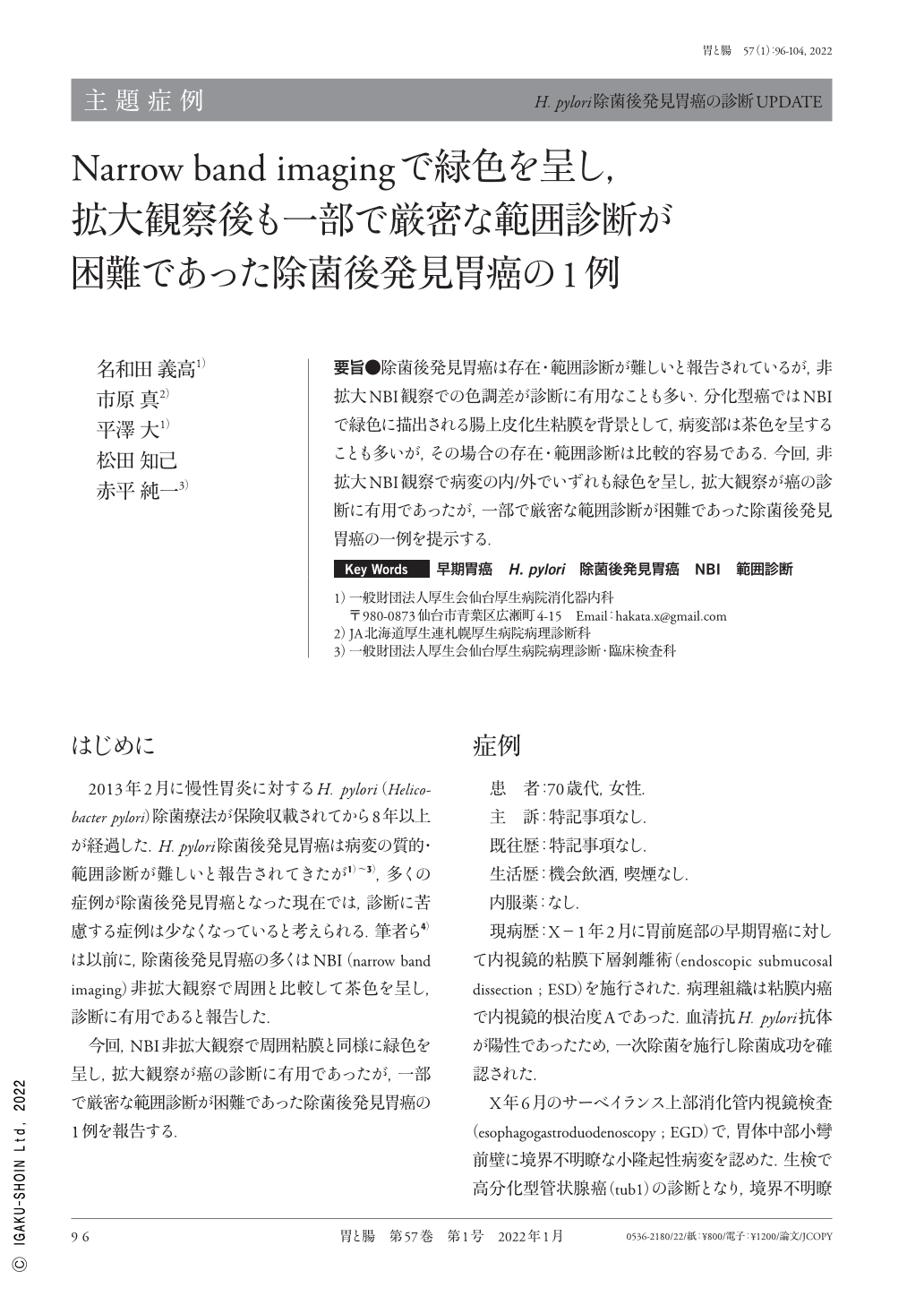
要旨●除菌後発見胃癌は存在・範囲診断が難しいと報告されているが,非拡大NBI観察での色調差が診断に有用なことも多い.分化型癌ではNBIで緑色に描出される腸上皮化生粘膜を背景として,病変部は茶色を呈することも多いが,その場合の存在・範囲診断は比較的容易である.今回,非拡大NBI観察で病変の内/外でいずれも緑色を呈し,拡大観察が癌の診断に有用であったが,一部で厳密な範囲診断が困難であった除菌後発見胃癌の一例を提示する.
It has been reported that the presence and extent of gastric cancer occurring after Helicobacter pylori eradication is difficult to diagnose, but the color difference in nonmagnified NBI(narrow band imaging)is often useful for diagnosis. In the case of differentiated tubular adenocarcinoma, the lesion often appears brownish against the background of the intestinal metaplasia mucosa, which is depicted as greenish in NBI, and the diagnosis of the presence and extent of the lesion is relatively easy. We present a case of gastric cancer detected after eradication in which the inner and outer sides of the lesion were both greenish on nonmagnified NBI. Although magnified observation was useful in diagnosing the cancer, strict boundary diagnosis was difficult in some areas.
Copyright © 2022, Igaku-Shoin Ltd. All rights reserved.