Japanese
English
- 有料閲覧
PPV購入案内
1,320円 (1,200円+税10%) こちらは、1058ページから1062ページの文献です。購入には医書.jp本会員登録が必要です。
会員登録完了後に改めて上記「購入サイトへ」を選択してください。
または「購入サイトへ」選択後に「購入ログイン」、「新規会員登録」の順に進んでください。
会員登録について詳しくはをご確認ください。
- Abstract 文献概要
- 1ページ目 Look Inside
- 参考文献 Reference
- サイト内被引用 Cited by
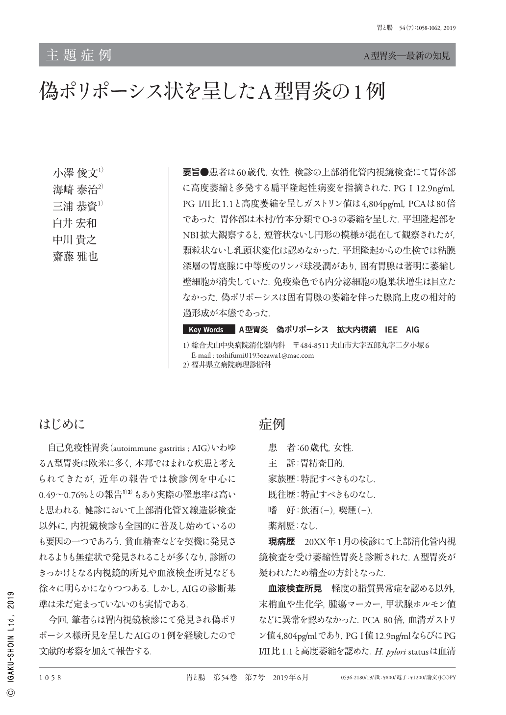
要旨●患者は60歳代,女性.検診の上部消化管内視鏡検査にて胃体部に高度萎縮と多発する扁平隆起性病変を指摘された.PG I 12.9ng/ml,PG I/II比1.1と高度萎縮を呈しガストリン値は4,804pg/ml,PCAは80倍であった.胃体部は木村/竹本分類でO-3の萎縮を呈した.平坦隆起部をNBI拡大観察すると,短管状ないし円形の模様が混在して観察されたが,顆粒状ないし乳頭状変化は認めなかった.平坦隆起からの生検では粘膜深層の胃底腺に中等度のリンパ球浸潤があり,固有胃腺は著明に萎縮し壁細胞が消失していた.免疫染色でも内分泌細胞の胞巣状増生は目立たなかった.偽ポリポーシスは固有胃腺の萎縮を伴った腺窩上皮の相対的過形成が本態であった.
A female in her 60s was admitted to our department for further examination of atrophic gastritis. Her antiparietal cell antibody level was 80 times the normal level, and her serum gastrin level was 4,804pg/ml. Her pepsinogen(PG)-I level was 12.9ng/ml, and the PG-I/PG-II ratio was 1.1, suggesting severe atrophy. Gastrography showed numerous polypoid lesions of the gastric body and fornix. Gastroscopy showed severely atrophic mucosa(Kimura-Takemoto Classification:O-3)at the gastric body and fornix, except for the antrum. After spraying indigo carmine dye, we noted numerous flat elevated lesions, which were approximately the same size. NBI-magnifying endoscopy showed round and short-tubular structures on flat elevated lesions. The biopsy specimens of the elevated lesions revealed atrophic proper gastric glands and totally extinguished parietal cells. Endocrine micronest has not been proven despite additional immunostaining. In this patient, pseudopolyposis was caused by hypertrophy of the foveolar epithelium with atrophic proper gastric glands, not NET or collagenous gastritis.
Copyright © 2019, Igaku-Shoin Ltd. All rights reserved.