Japanese
English
- 有料閲覧
- Abstract 文献概要
- 1ページ目 Look Inside
- 参考文献 Reference
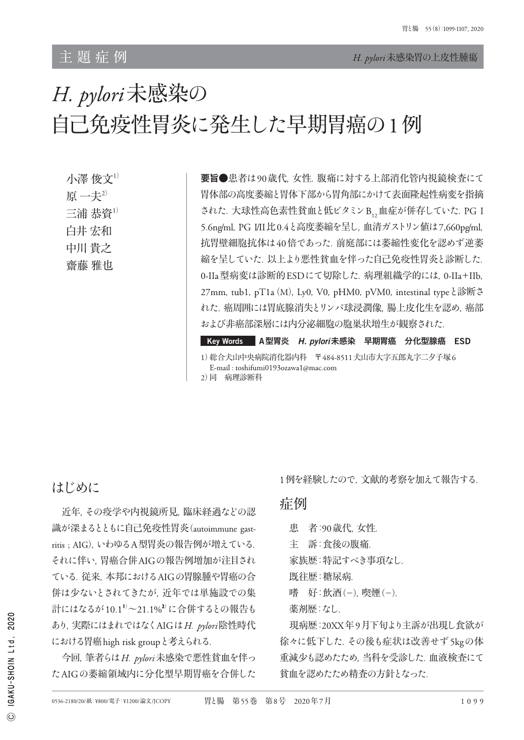
要旨●患者は90歳代,女性.腹痛に対する上部消化管内視鏡検査にて胃体部の高度萎縮と胃体下部から胃角部にかけて表面隆起性病変を指摘された.大球性高色素性貧血と低ビタミンB12血症が併存していた.PG I 5.6ng/ml,PG I/II比0.4と高度萎縮を呈し,血清ガストリン値は7,660pg/ml,抗胃壁細胞抗体は40倍であった.前庭部には萎縮性変化を認めず逆萎縮を呈していた.以上より悪性貧血を伴った自己免疫性胃炎と診断した.0-IIa型病変は診断的ESDにて切除した.病理組織学的には,0-IIa+IIb,27mm,tub1,pT1a(M),Ly0,V0,pHM0,pVM0,intestinal typeと診断された.癌周囲には胃底腺消失とリンパ球浸潤像,腸上皮化生を認め,癌部および非癌部深層には内分泌細胞の胞巣状増生が観察された.
A woman in her 90s with abdominal discomfort was admitted to our department for further examination. She had pernicious anemia alongside vitamin B12 deficiency. Her anti-parietal cell antibody level was 40 times and her serum gastrin level was 7,660pg/ml. Her PG(pepsinogen)-I level was 5.6ng/ml and the I/II ratio was 0.4, therefore severe atrophy was assumed. Gastroscopy showed severe atrophic mucosa(Kimura-Takemoto Classification:O-3)at the gastric body and fornix except for the antrum. The patient was diagnosed with autoimmune gastritis. Furthermore, a flat elevated lesion 25mm in diameter was detected endoscopically at the lower gastric body covered with atrophic mucosa. This lesion was diagnosed as an early gastric mucosal cancer and resected by ESD. Macroscopic type was 0-IIa+IIb and histological examination showed well differentiated adenocarcinoma, pT1a(M), with intestinal type mucin phenotype. Analysis of the resected specimen revealed proper gastric glands and parietal cells totally extinguished, with widespread lymphocyte infiltration. Endocrine micronests which were positive for chromogranin A staining were observed in the deeper mucosal layer of both the cancerous and non-cancerous area with intestinal metaplasia.

Copyright © 2020, Igaku-Shoin Ltd. All rights reserved.


