Japanese
English
- 有料閲覧
PPV購入案内
1,320円 (1,200円+税10%) こちらは、105ページから112ページの文献です。購入には医書.jp本会員登録が必要です。
会員登録完了後に改めて上記「購入サイトへ」を選択してください。
または「購入サイトへ」選択後に「購入ログイン」、「新規会員登録」の順に進んでください。
会員登録について詳しくはをご確認ください。
- Abstract 文献概要
- 1ページ目 Look Inside
- 参考文献 Reference
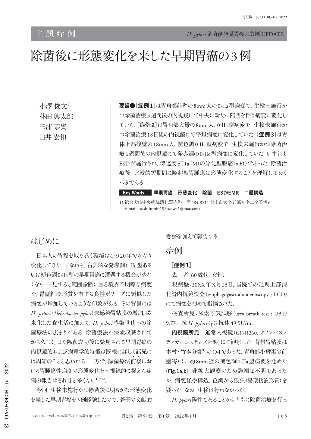
要旨●[症例1]は胃角部前壁の8mm大の0-IIa型病変で,生検未施行かつ除菌治療3週間後の内視鏡にて中央に新たに陥凹を伴う病変に変化していた.[症例2]は胃角部大彎の8mm大,0-IIa型病変で,生検未施行かつ除菌治療18日後の内視鏡にて平坦病変に変化していた.[症例3]は胃体上部後壁の18mm大,褪色調0-IIa型病変で,生検未施行かつ除菌治療6週間後の内視鏡にて発赤調の0-IIc型病変に変化していた.いずれもESDが施行され,深達度pT1a(M)の分化型腺癌(tub1)であった.除菌治療後,比較的短期間に隆起型胃腫瘍は形態変化することを理解しておくべきである.
Case 1 was a female in her 60s who underwent EGD(esophagogastroduodenoscopy)during a health checkup, which detected an elevated lesion 5mm in diameter located on the anterior wall of the gastric angle. An EGD performed three weeks after Helicobacter pylori eradication therapy revealed that this lesion had a depressed area in its center. ESD(Endoscopic submucosal dissection)was continued, and histopathological examination showed a mucosal adenocarcinoma(tub1), type 0-IIa+IIc.
Case 2 was a male in his 70s who underwent EGD during a health checkup, which detected an elevated lesion 8mm in diameter located at the greater curvature of the gastric angle. An EGD performed 18 days after H. pylori eradication therapy revealed that this lesion had changed from an elevated lesion into a flat lesion. ESD was continued, and histopathological examination showed a mucosal adenocarcinoma(tub1), type 0-IIa.
Case 3 was a male in his 60s who underwent EGD during a health checkup, which detected an elevated lesion 18 mm in diameter, located on the posterior wall of the upper gastric body. An EGD performed six weeks after H. pylori eradication therapy revealed that this lesion had changed into a reddish depressed lesion. ESD was continued, and histopathological examination showed a mucosal adenocarcinoma(tub1), type 0-IIc. All three cases had never undergone biopsy.
We concluded that morphological changes can occur in gastric tumors after H. pylori eradication therapy.
Copyright © 2022, Igaku-Shoin Ltd. All rights reserved.