Japanese
English
- 有料閲覧
PPV購入案内
1,320円 (1,200円+税10%) こちらは、1684ページから1692ページの文献です。購入には医書.jp本会員登録が必要です。
会員登録完了後に改めて上記「購入サイトへ」を選択してください。
または「購入サイトへ」選択後に「購入ログイン」、「新規会員登録」の順に進んでください。
会員登録について詳しくはをご確認ください。
- Abstract 文献概要
- 1ページ目 Look Inside
- 参考文献 Reference
- サイト内被引用 Cited by
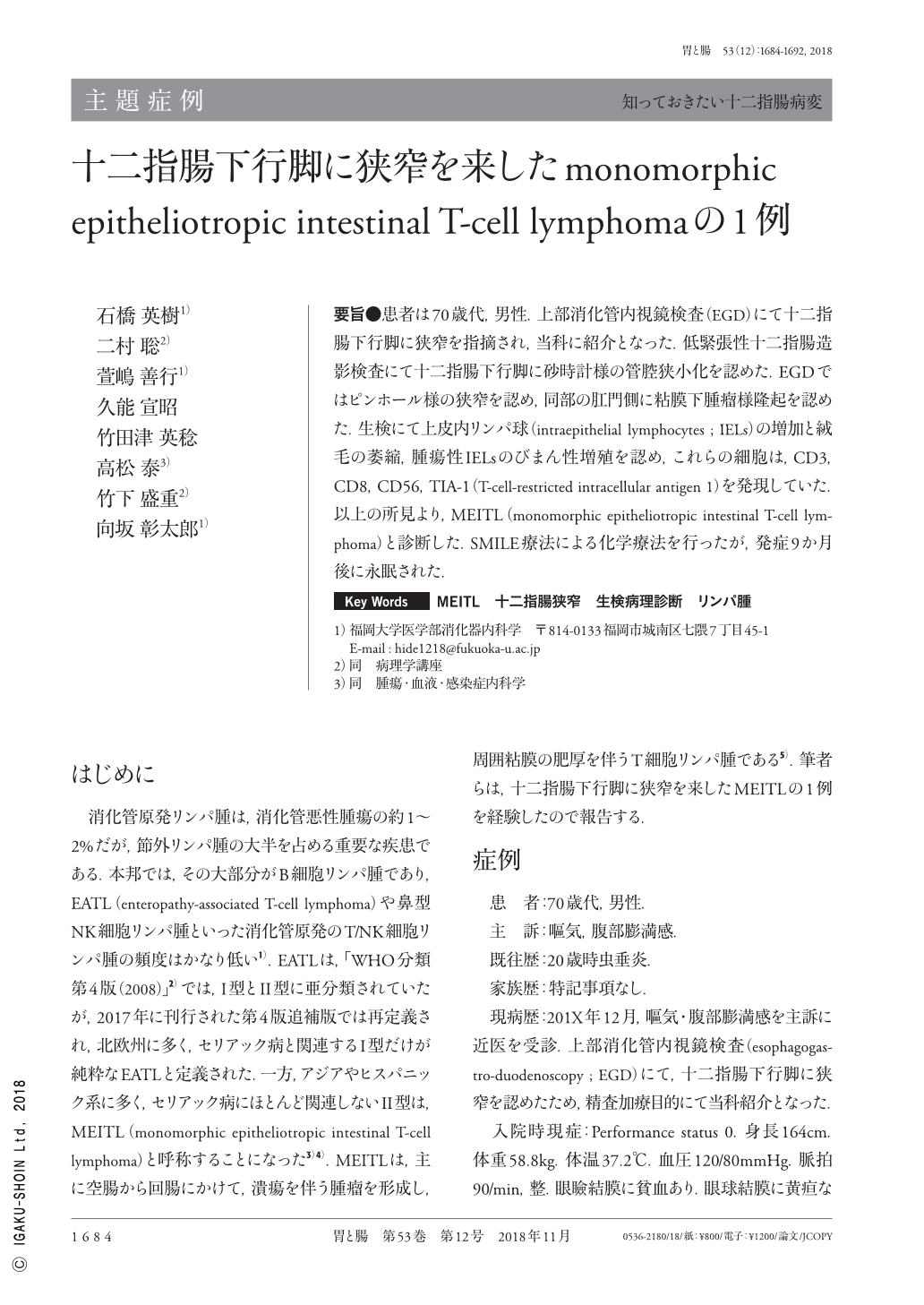
要旨●患者は70歳代,男性.上部消化管内視鏡検査(EGD)にて十二指腸下行脚に狭窄を指摘され,当科に紹介となった.低緊張性十二指腸造影検査にて十二指腸下行脚に砂時計様の管腔狭小化を認めた.EGDではピンホール様の狭窄を認め,同部の肛門側に粘膜下腫瘤様隆起を認めた.生検にて上皮内リンパ球(intraepithelial lymphocytes ; IELs)の増加と絨毛の萎縮,腫瘍性IELsのびまん性増殖を認め,これらの細胞は,CD3,CD8,CD56,TIA-1(T-cell-restricted intracellular antigen 1)を発現していた.以上の所見より,MEITL(monomorphic epitheliotropic intestinal T-cell lymphoma)と診断した.SMILE療法による化学療法を行ったが,発症9か月後に永眠された.
A 70-year-old man was admitted for nausea. Upper GI endoscopy revealed severe pinhole-like stenosis, a submucosal tumor on the anal side of the stenosis, and edematous mucosa in the second portion of the duodenum. Lower GI endoscopy revealed granular mucosa of the terminal ileum and edematous or normal-looking colorectal mucosa. Duodenitis with villous atrophy and abundant IELs(intraepithelial lymphocytes)was evident in the biopsy specimens from the second portion of the duodenum taken outside the tumor area. The tumor itself comprised small- to medium-sized atypical lymphocytes both among the epithelial cells and in the mucosal layer. The cecal mucosa was characterized by severe infiltrate of atypical IELs. Other biopsy specimens from the duodenum, descending colon, and rectum revealed chronic inflammatory changes with increased CD3- and CD8-positive and CD56-negative T-IELs. Therefore, we diagnosed monomorphic epitheliotropic intestinal T-cell lymphoma. The patient was treated with a chemotherapy regimen of SMILE. Post chemotherapy, abdominal CT revealed a reduction in the tumor size. Despite a partial response to chemotherapy, he died of sepsis nine months after the disease onset.
Copyright © 2018, Igaku-Shoin Ltd. All rights reserved.