Japanese
English
- 有料閲覧
PPV購入案内
1,320円 (1,200円+税10%) こちらは、52ページから63ページの文献です。購入には医書.jp本会員登録が必要です。
会員登録完了後に改めて上記「購入サイトへ」を選択してください。
または「購入サイトへ」選択後に「購入ログイン」、「新規会員登録」の順に進んでください。
会員登録について詳しくはをご確認ください。
- Abstract 文献概要
- 1ページ目 Look Inside
- 参考文献 Reference
- サイト内被引用 Cited by
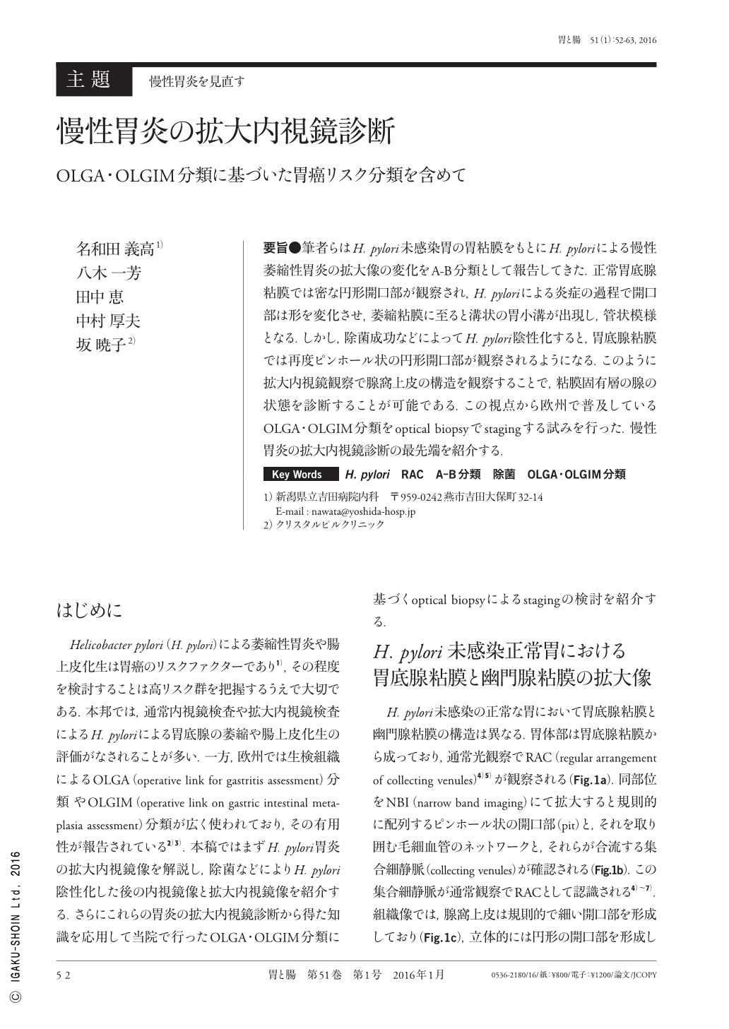
要旨●筆者らはH. pylori未感染胃の胃粘膜をもとにH. pyloriによる慢性萎縮性胃炎の拡大像の変化をA-B分類として報告してきた.正常胃底腺粘膜では密な円形開口部が観察され,H. pyloriによる炎症の過程で開口部は形を変化させ,萎縮粘膜に至ると溝状の胃小溝が出現し,管状模様となる.しかし,除菌成功などによってH. pylori陰性化すると,胃底腺粘膜では再度ピンホール状の円形開口部が観察されるようになる.このように拡大内視鏡観察で腺窩上皮の構造を観察することで,粘膜固有層の腺の状態を診断することが可能である.この視点から欧州で普及しているOLGA・OLGIM分類をoptical biopsyでstagingする試みを行った.慢性胃炎の拡大内視鏡診断の最先端を紹介する.
We have previously defined and reported the A-B classification, which illustrates the transformation of the surface structure in a Helicobacter pylori(H. pylori)-positive stomach via ME(magnifying endoscopy). Round pits are observed in the fundic gland mucosa in H. pylori-negative normal stomachs on ME. However, the surface epithelium structure changes during inflammation caused by H. pylori, and sulci are observed in the atrophic mucosa on ME. Both round pits and sulci correspond to crypts. After successful eradication, pinhole pits may be observed in the fundic gland. The mucosa that develops in the fundic gland, even when the mucosa was atrophic and had no fundic glands before, shows round pits. As in these descriptions, the surface epithelium of the stomach changes its structure due to inflammation and tends to return to the structure of a normal mucosa after eradication of H. pylori. Applying this rationale, we examined whether ME would be as useful as optical biopsy for the OLGA/OLGIM staging system by comparing ME images with microscopic images of biopsy specimens taken from the corresponding area. The stage concordance rate between ME and the histological images was relatively high, and the application of the optical biopsy using ME to the OLGA/OLGIM staging system was considered useful to some extent.
Copyright © 2016, Igaku-Shoin Ltd. All rights reserved.