Japanese
English
- 有料閲覧
- Abstract 文献概要
- 1ページ目 Look Inside
- 参考文献 Reference
- サイト内被引用 Cited by
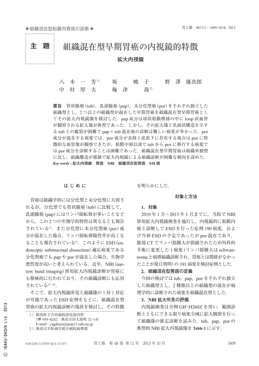
要旨 管状腺癌(tub),乳頭腺癌(pap),未分化型癌(por)をそれぞれ独立した組織型とし,2つ以上の組織型が混在した早期胃癌を組織混在型早期胃癌としてその拡大内視鏡像を検討した.pap成分は球状粘膜模様の中にloop状血管が観察される拡大像が典型であった.しかし,その拡大像と乳頭状構造を呈するtubとの鑑別が困難でpap+tub混在癌の診断は難しい病変が多かった.por成分が混在する病変では,por成分が表層上皮直下に存在する場合はporに特徴的な血管像が観察できたが,粘膜中層以深でtubからporに移行する病変ではpor成分を診断することは困難であった.組織混在型早期胃癌は組織単独型に比し,組織構造が複雑で拡大内視鏡による組織診断が困難な傾向を認めた.
We studied the magnifying endoscopic findings of a histological mixed type of gastric adenocarcinoma, knowing that tubular adenocarcinoma(tub), papillary adenocarcinoma(pap)and undifferentiated adenocarcinoma(por)are independent histological types and two or three mixed-type are regarded as histological mixed type. The typical magnifying endoscopic finding of pap is a sphere-shaped pattern with loop-like vascular vessels. However, it is difficult to distinguish this magnifying endoscopic finding from tub with a papillary structure, so, the mixed-type adenocarcinoma of tub and pap is difficult to diagnose by magnifying endoscopy. In the case of mixed type of tub and por, if the component of por exists beneath the surface layer of the epithelium, it is not difficult to recognize because the typical magnifying endoscopic finding can be seen. However, if por is transformed from tub in a deep layer of the mucosa, the component of por cannot be diagnosed by magnifying endoscopy. The histological mixed-type is complex in its histological structure, so its magnifying endoscopic diagnosis is thought to be difficult compared with the histological single type of gastric adenocarcinoma.

Copyright © 2013, Igaku-Shoin Ltd. All rights reserved.


