Japanese
English
- 有料閲覧
- Abstract 文献概要
- 1ページ目 Look Inside
- 参考文献 Reference
- サイト内被引用 Cited by
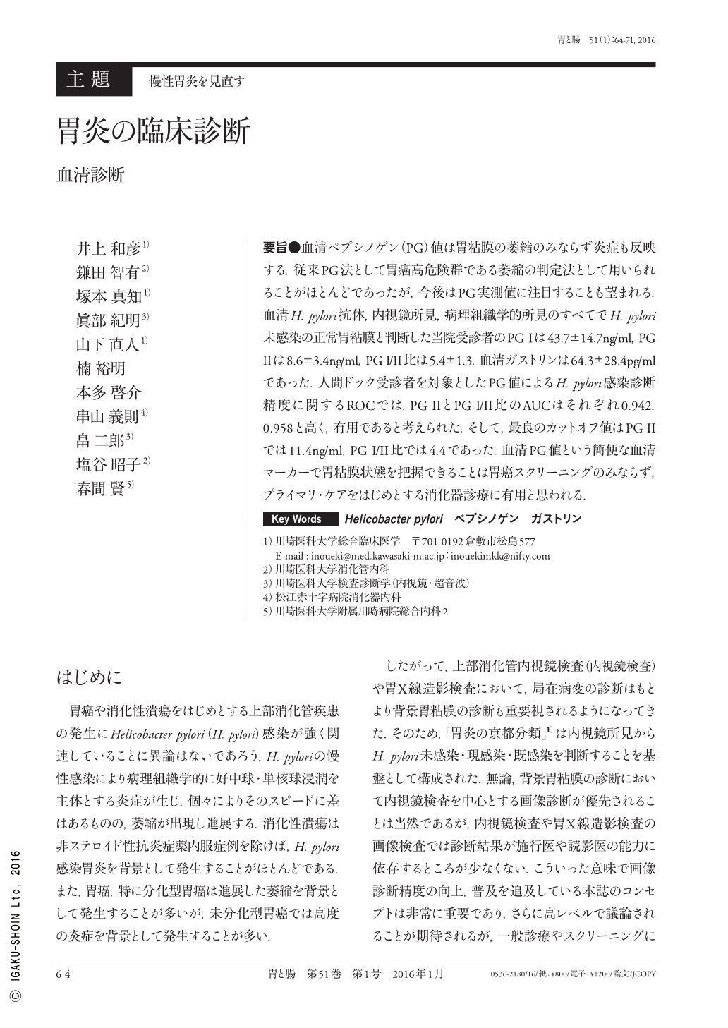
要旨●血清ペプシノゲン(PG)値は胃粘膜の萎縮のみならず炎症も反映する.従来PG法として胃癌高危険群である萎縮の判定法として用いられることがほとんどであったが,今後はPG実測値に注目することも望まれる.血清H. pylori抗体,内視鏡所見,病理組織学的所見のすべてでH. pylori未感染の正常胃粘膜と判断した当院受診者のPG Iは43.7±14.7ng/ml,PG IIは8.6±3.4ng/ml,PG I/II比は5.4±1.3,血清ガストリンは64.3±28.4pg/mlであった.人間ドック受診者を対象としたPG値によるH. pylori感染診断精度に関するROCでは,PG IIとPG I/II比のAUCはそれぞれ0.942,0.958と高く,有用であると考えられた.そして,最良のカットオフ値はPG IIでは11.4ng/ml,PG I/II比では4.4であった.血清PG値という簡便な血清マーカーで胃粘膜状態を把握できることは胃癌スクリーニングのみならず,プライマリ・ケアをはじめとする消化器診療に有用と思われる.
Serum pepsinogen(PG)levels reflect not only gastric atrophy but also levels of inflammation. Although PG levels have primarily been utilized for the detection of atrophy that has a high risk of evolving into gastric cancer, focusing on PG measurements may provide other benefits in the future. Patients who visited our hospital and were diagnosed with normal gastric mucosa without Helicobacter pylori infection, based on three tests(serum anti-H. pylori antibody levels, endoscopy, and histology), had PG I levels of 43.7±14.7ng/mL, PG II levels of 8.6±3.4ng/mL, a PG I/II ratio of 5.4±1.3, and serum gastrin levels of 64.3±28.4pg/mL. In the health checkup participants, the areas under the receiver operating characteristic curve for the diagnosis of H. pylori infection using the PG II levels and PG I/II ratio were as high as 0.942 and 0.958, respectively ; this indicated their advantages. The optimal cut-offs for the PG II levels and PG I/II ratio were 11.4ng/mL and 4.4, respectively. A simple serologic marker to capture the status of the gastric mucosa would be useful not only in the screening for gastric cancer but also for management of gastrointestinal disorders, including primary care.

Copyright © 2016, Igaku-Shoin Ltd. All rights reserved.


