Japanese
English
- 有料閲覧
PPV購入案内
1,320円 (1,200円+税10%) こちらは、406ページから410ページの文献です。購入には医書.jp本会員登録が必要です。
会員登録完了後に改めて上記「購入サイトへ」を選択してください。
または「購入サイトへ」選択後に「購入ログイン」、「新規会員登録」の順に進んでください。
会員登録について詳しくはをご確認ください。
- Abstract 文献概要
- 1ページ目 Look Inside
- 参考文献 Reference
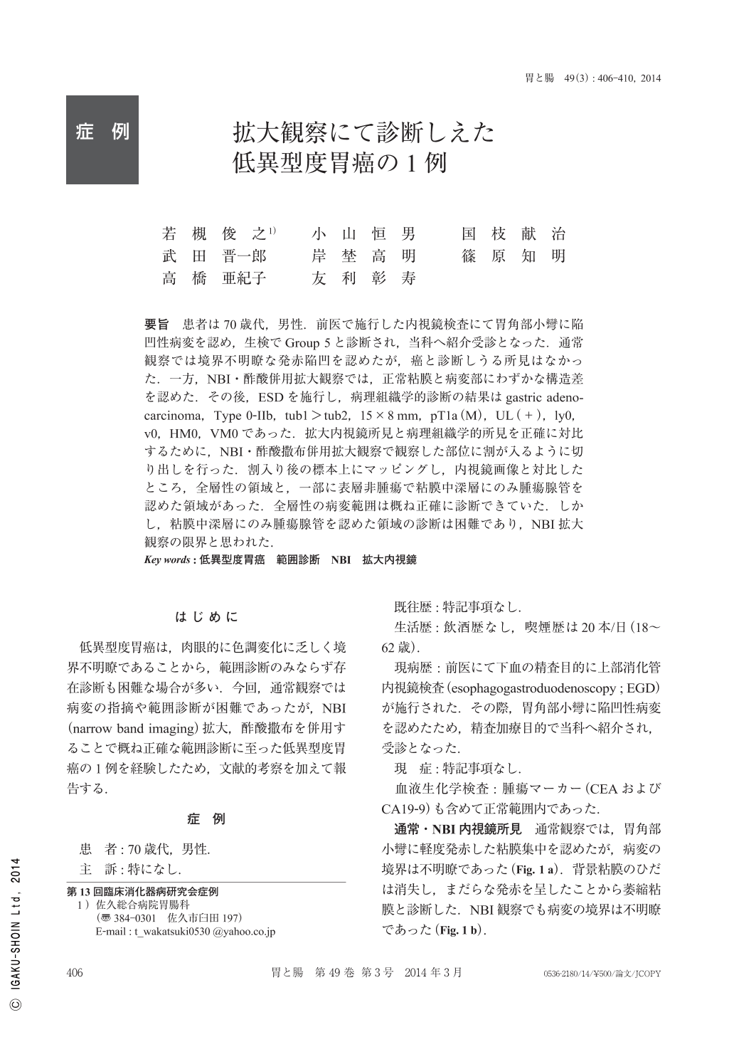
要旨 患者は70歳代,男性.前医で施行した内視鏡検査にて胃角部小彎に陥凹性病変を認め,生検でGroup 5と診断され,当科へ紹介受診となった.通常観察では境界不明瞭な発赤陥凹を認めたが,癌と診断しうる所見はなかった.一方,NBI・酢酸併用拡大観察では,正常粘膜と病変部にわずかな構造差を認めた.その後,ESDを施行し,病理組織学的診断の結果はgastric adenocarcinoma,Type 0-IIb,tub1>tub2,15×8mm,pT1a(M),UL(+),ly0,v0,HM0,VM0であった.拡大内視鏡所見と病理組織学的所見を正確に対比するために,NBI・酢酸撒布併用拡大観察で観察した部位に割が入るように切り出しを行った.割入り後の標本上にマッピングし,内視鏡画像と対比したところ,全層性の領域と,一部に表層非腫瘍で粘膜中深層にのみ腫瘍腺管を認めた領域があった.全層性の病変範囲は概ね正確に診断できていた.しかし,粘膜中深層にのみ腫瘍腺管を認めた領域の診断は困難であり,NBI拡大観察の限界と思われた.
A 70s-year-old man was referred to our hospital for work up of a gastric lesion diagnosed as cancer by biopsy. A reddish lesion was found at the angle. Because the margin was unclear in white light imaging, it was diagnosed as gastritis. However, ME(magnified endoscopy)with NBI and acetic acid revealed a slight irregular villous pattern in the lesion. Lateral extension was diagnosed by the difference in surface pattern. En block ESD(endoscopic submucosal dissection)was performed, and pathological diagnosis was well differentiated adenocarcinoma, Type 0-IIb, tub1>tub2, 15×8mm, pT1a(M), UL(+), ly0, v0, HM0, VM0.
The center of the lesion was cut to compare ME and histological findings.
Cancer exposed to the surface in the central part of the lesion, and was diagnosed correctly. However, it was partially covered by non-neoplastic epithelium in the peripheral area, and was not diagnosed by ME. Diagnosing such cases of buried adenocarcinoma is beyond NBI's ability.
Copyright © 2014, Igaku-Shoin Ltd. All rights reserved.