Japanese
English
- 有料閲覧
PPV購入案内
1,320円 (1,200円+税10%) こちらは、781ページから787ページの文献です。購入には医書.jp本会員登録が必要です。
会員登録完了後に改めて上記「購入サイトへ」を選択してください。
または「購入サイトへ」選択後に「購入ログイン」、「新規会員登録」の順に進んでください。
会員登録について詳しくはをご確認ください。
- Abstract 文献概要
- 1ページ目 Look Inside
- 参考文献 Reference
- サイト内被引用 Cited by
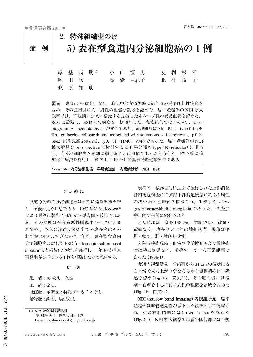
要旨 患者は70歳代,女性.胸部中部食道後壁に褪色調の扁平隆起性病変を認め,その肛門側に約半周性の粗糙な領域を認めた.扁平隆起部のNBI拡大観察では,不規則に分岐・横走する拡張した非ループ性の異常血管を認めた.SCCと診断し,ESDにて病変を一括切除した.免疫染色ではN-CAM,chromogranin A,synaptophysinが陽性であり,病理診断はMt,Post,type 0-IIa+IIb,endocrine cell carcinoma associated with squamous cell carcinoma,pT1b-SM2(浸潤距離250μm),ly0,v1,HM0,VM0であった.扁平隆起部のNBI拡大所見をretrospectiveに検討すると有馬分類のtype 4R(reticular)に相当し,内分泌細胞癌を鑑別に挙げることは可能であったと考えた.ESD後に追加化学療法を施行し,術後1年10か月間無再発経過観察中である.
The patient was a female in her seventies. Conventional endoscopy showed a discolored flat elevated lesion and a white flat lesion with coarse surface on the posterior wall of the middle thoracic esophagus.
NBI(narrow band imaging)magnified endoscopy revealed irregularly-branched and dilated non-loop vessels on the surface of the flat elevated lesion.
We performed ESD(endoscopic submucosal dissection)for the lesion in en-bloc fashion. Histologically, the tumor was diagnosed as an esophageal endocrine cell carcinoma associated with Mt, Post, type 0-IIa+IIb, squamous cell carcinoma, pT1b-SM2(invasive depth 250μm), ly0, v1, HM0, VM0.
Immmunohistochemically, N-CAM, chromogranin A and synaptophysin were positive for the tumor cells.
The microvascular pattern of the flat elevated lesion was judged as being the reticular type. Sometimes, this type of irregular microvascular pattern was observed in undifferentiated SCC(squamous cell carcinoma). The patient was treated with adjuvant chemotherapy after ESD and is still living without recurrence 22 months later.
Copyright © 2011, Igaku-Shoin Ltd. All rights reserved.