Japanese
English
- 有料閲覧
PPV購入案内
1,320円 (1,200円+税10%) こちらは、1360ページから1368ページの文献です。購入には医書.jp本会員登録が必要です。
会員登録完了後に改めて上記「購入サイトへ」を選択してください。
または「購入サイトへ」選択後に「購入ログイン」、「新規会員登録」の順に進んでください。
会員登録について詳しくはをご確認ください。
- Abstract 文献概要
- 1ページ目 Look Inside
- 参考文献 Reference
- サイト内被引用 Cited by
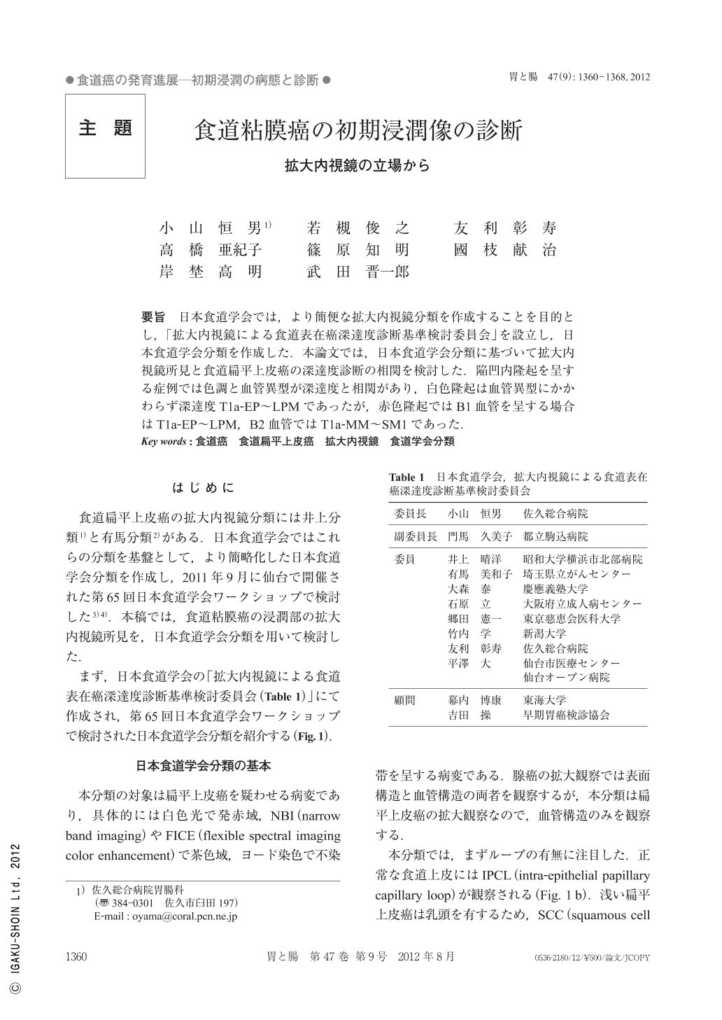
要旨 日本食道学会では,より簡便な拡大内視鏡分類を作成することを目的とし,「拡大内視鏡による食道表在癌深達度診断基準検討委員会」を設立し,日本食道学会分類を作成した.本論文では,日本食道学会分類に基づいて拡大内視鏡所見と食道扁平上皮癌の深達度診断の相関を検討した.陥凹内隆起を呈する症例では色調と血管異型が深達度と相関があり,白色隆起は血管異型にかかわらず深達度T1a-EP~LPMであったが,赤色隆起ではB1血管を呈する場合はT1a-EP~LPM,B2血管ではT1a-MM~SM1であった.
A new classification of magnified endoscopic classification of superficial esophageal SCC(squamous cell carcinoma)was established by the Japan esophageal association. The micro vascular pattern was classified into B1, B2 and B3. B1 : loop like vessels. B2 : non-loop vessels. B3 : Vessels three times or more thicker than B2. There are some cases with elevation within flat depressed lesion(0-IIc+IIa)invading the MM(muscularis mucosae). A white elevated area doesn't invade the Muscularis mucosae regardless of vascular pattern. However, a red elevated area with B2 vascular pattern invaded the MM or SM1. Therefore, this new classification of the Japan Esophageal Association is useful for the detection of the minute invasion site of superficial esophageal SCC.
Copyright © 2012, Igaku-Shoin Ltd. All rights reserved.