Japanese
English
- 有料閲覧
PPV購入案内
1,320円 (1,200円+税10%) こちらは、555ページから560ページの文献です。購入には医書.jp本会員登録が必要です。
会員登録完了後に改めて上記「購入サイトへ」を選択してください。
または「購入サイトへ」選択後に「購入ログイン」、「新規会員登録」の順に進んでください。
会員登録について詳しくはをご確認ください。
- Abstract 文献概要
- 1ページ目 Look Inside
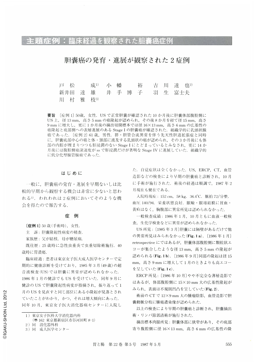
要旨 〔症例1〕50歳,女性.USで正常胆囊が確認された10か月後に胆囊体部腹腔側にUS上,径13mm,高さ5mmの癌隆起が認められ,その後8か月を経て径15mm,高さ9mmに増大し,更に1か月後の摘出切開標本では径16×13mm,高さ6mmの広基性の癌隆起と底部側への表層進展のあるStage Ⅰの胆囊癌が確認された.組織学的に乳頭状腺癌であった.〔症例2〕61歳,男性.膵・胆管合流異常を伴う先天性胆道拡張症と同時に,胆囊底部中心の癌と体・頸部に波及する乳頭状の癌が認められ,その3か月後にも体部の内腔が埋まりつつも肝浸潤のないStage Ⅰにとどまっているとみなされ,更に14か月後には腹腔側癌深達度がssで肝浸潤だけが著明なStage Ⅳに進展していた.組織学的に低分化型腺管腺癌であった.
We observed the growth and extension of early or relatively-early gallbladder cancer in two patients.
〔Case 1〕 50 year-old female: Ten months after the patient was confirmed to have no abnormality in the gallbladder by ultrasonography. A protruding lesion 13 mm in diameter and 5 mm in height was detected on the peritoneal side of the corpus by ultrasonography. Eight months after that, it had grown to 15 mm in diameter and 9 mm in height. Operation was performed one month afterwards. Examination of the excised and incised gallbladder material revealed a broad-based protruding tumor 16×13 mm in diameters and 6 mm in height extending superficially towards the fundus, which was identified as Stage Ⅰ carcinoma of the gallbladder. Histologically, it was papillary adenocarcinoma.
〔Case 2〕 61 year-old male: Coincidentally with an anomalous pancreaticobiliary ductal system, a cancer at the gallbladder fundus and a papillary cancer spreading to the corpus and neck were detected. Three months after that, the lesion seemed to have remained at Stage Ⅰ, as there was no clinical infiltration though the lumen of the corpus was going to be filled. Fourteen months after that, the lesion had progressed to Stage Ⅳ, penetrating to the depth of the peritoneal side of the gallbladder and markedly infiltrating the liver. Histologically, it was poorly differentiated tubular adenocarcinoma.
Copyright © 1987, Igaku-Shoin Ltd. All rights reserved.