Japanese
English
- 有料閲覧
- Abstract 文献概要
- 1ページ目 Look Inside
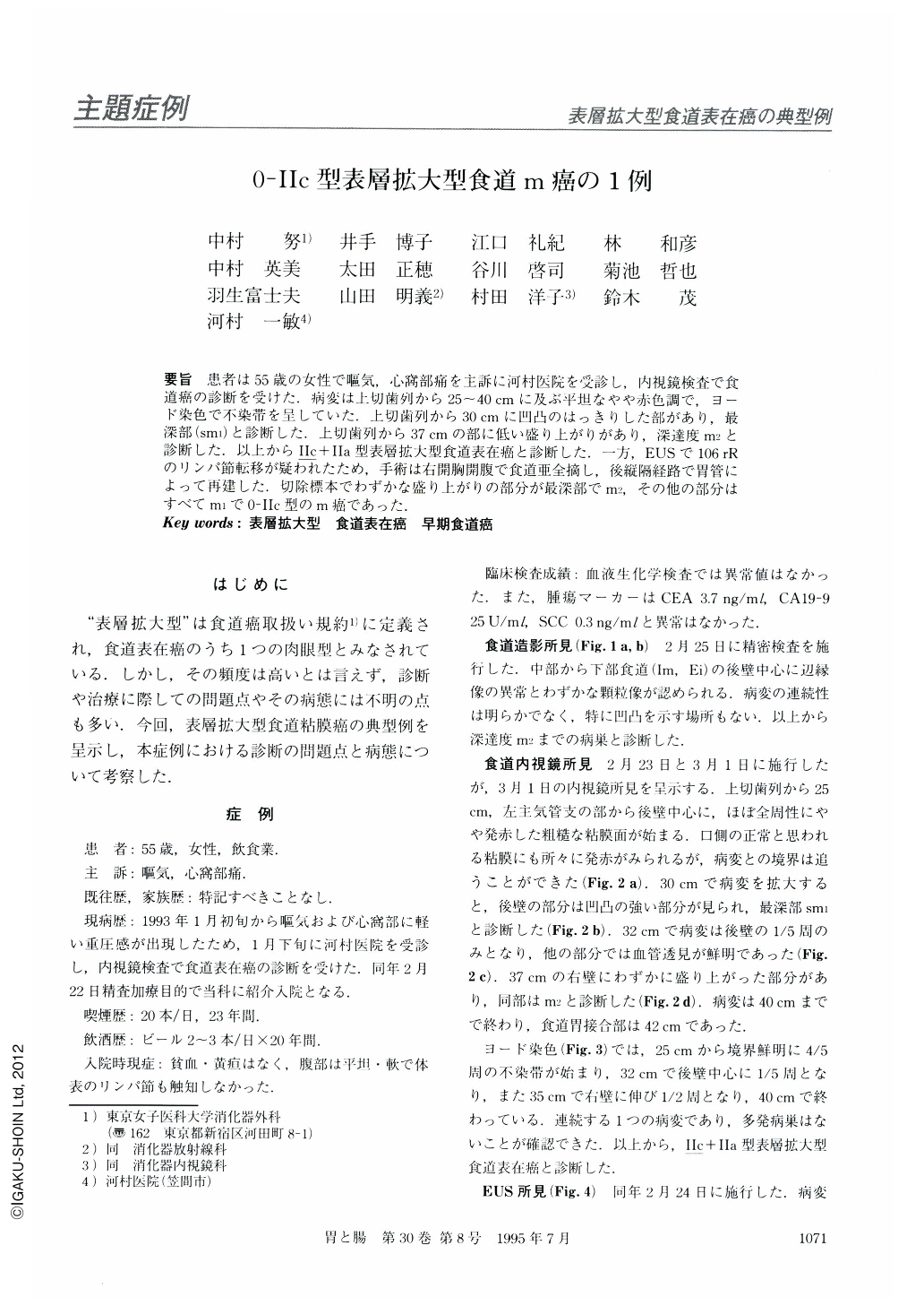
要旨 患者は55歳の女性で嘔気,心窩部痛を主訴に河村医院を受診し,内視鏡検査で食道癌の診断を受けた.病変は上切歯列から25~40cmに及ぶ平坦なやや赤色調で,ヨード染色で不染帯を呈していた.上切歯列から30cmに凹凸のはっきりした部があり,最深部(SM1)と診断した.上切歯列から37cmの部に低い盛り上がりがあり,深達度m2と診断した.以上からⅡc+Ⅱa型表層拡大型食道表在癌と診断した.一方,EUSで106rRのリンパ節転移が疑われたため,手術は右開胸開腹で食道亜全摘し,後縦隔経路で胃管によって再建した.切除標本でわずかな盛り上がりの部分が最深部でm2,その他の部分はすべてm1で0-Ⅱc型のm癌であった.
A 55-year-old female with a complaint of nausea and epigastralgia was diagnosed by endoscopy as having esophageal cancer. Her lesion was an area (0-Ⅱc) of slight redness without iodine staining which extended between 25 cm and 40 cm from the incisors. Depth of invasion in the depressed lesion at 30 cm was diagnosed as sm1 cancer and that in the low protuberant lesion at 37 cm was diagnosed as m2. EUS findings showed swelling of the right reccurent nerve nodes (106 rR). The patient underwent esophagectomy through right thoracotomy and reconstruction with a stomach tube.
Macroscopic feature of the lesion on the resected specimen was extended superficial depressed type (0-IIc type) cancer. Histologically, the deepest part of invasion, m2, was the low protuberant lesion in the lower esophagus.

Copyright © 1995, Igaku-Shoin Ltd. All rights reserved.


