Japanese
English
- 有料閲覧
PPV購入案内
1,320円 (1,200円+税10%) こちらは、961ページから967ページの文献です。購入には医書.jp本会員登録が必要です。
会員登録完了後に改めて上記「購入サイトへ」を選択してください。
または「購入サイトへ」選択後に「購入ログイン」、「新規会員登録」の順に進んでください。
会員登録について詳しくはをご確認ください。
- Abstract 文献概要
- 1ページ目 Look Inside
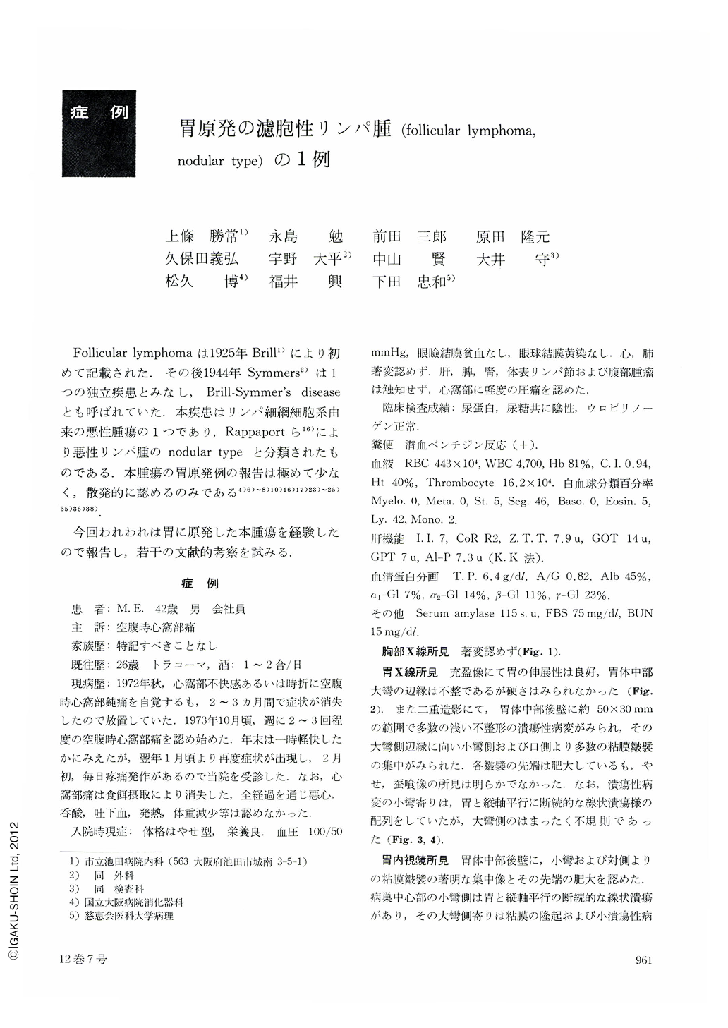
Follicular lymphomaは1925年Brill1)により初めて記載された.その後1944年Symmers2)は1つの独立疾患とみなし,Brill-Symmer's diseaseとも呼ばれていた,本疾患はリンパ細網細胞系由来の悪性腫瘍の1つであり,Rappaportら16)により悪性リンパ腫のnodular typeと分類されたものである.本腫瘍の胃原発例の報告は極めて少なく,散発的に認めるのみである4)6)~8)10)16)17)23)~25)35)36)38).
今回われわれは胃に原発した本腫瘍を経験したので報告し,若干の文献的考察を試みる.
The patient was a 42 year-old male, who had been admitted because of epigastric pain during the gastric emptying time.
The upper GI series showed numerous irregular and shallow ulcerous lesions accompanied with converging folds in an area of about 50×30 mm at the greater curvature side of the posterior wall of the middle corpus. Although the tip of each of the folds was enlarged, there was no definite sign of tapering nor moth-eaten appearance. And the gastric wall expanded well.
Endoscopically, the thickened mucosal folds looked like cerebral gyri, and the lesion showed multiple erosions mixed with marked nodular protuberances, but no definite margin of Ⅱc can be seen. The biopsy specimens revealed no sure sign of malignancy.
Gastrectomy was performed. No regional lymph node swelling was noted. In the excised specimen, multiple irregular ulcers were noted in the same area as was expected before. Although the tips of the convergent mucosal folds were enlarged and confluent, they were round and Ⅱc like lesion was not obvious. In addition, the mucosal surface showed partial nodular protuberance accompanied by bridging folds suspicious of submucosal tumor. From these findings, the ulcerous type of gastric lymphoma was highly suspected.
Histologically, the neoplastic change was observed in the submucosal area forming a submucosal ulcer (Ul-Ⅱ type of Murakami's classification). In the mucosa where proper gastric glands were destructed, there were infiltrations of mature lymphocytes. The submucosa was occupied by enlarged lymph follicles differing in size with conspicuous germinal center.
and some follicles were fused with oneanother. Lymphoid cells and reticulum cells in the follicles showed prominent pleomorphism. From these histological findings, this case was diagnosed as follicular lymphoma, nodular (“follicular”) mixed type (lymphocytic and reticulum cell) by Rappaport's classification.
It was fortunate that neoplastic tissue was limited to the submucosa and no signs of lymph node metastasis were observed. The prognosis is expected to be favorable.
Copyright © 1977, Igaku-Shoin Ltd. All rights reserved.