Japanese
English
- 有料閲覧
PPV購入案内
1,320円 (1,200円+税10%) こちらは、389ページから392ページの文献です。購入には医書.jp本会員登録が必要です。
会員登録完了後に改めて上記「購入サイトへ」を選択してください。
または「購入サイトへ」選択後に「購入ログイン」、「新規会員登録」の順に進んでください。
会員登録について詳しくはをご確認ください。
- Abstract 文献概要
- 1ページ目 Look Inside
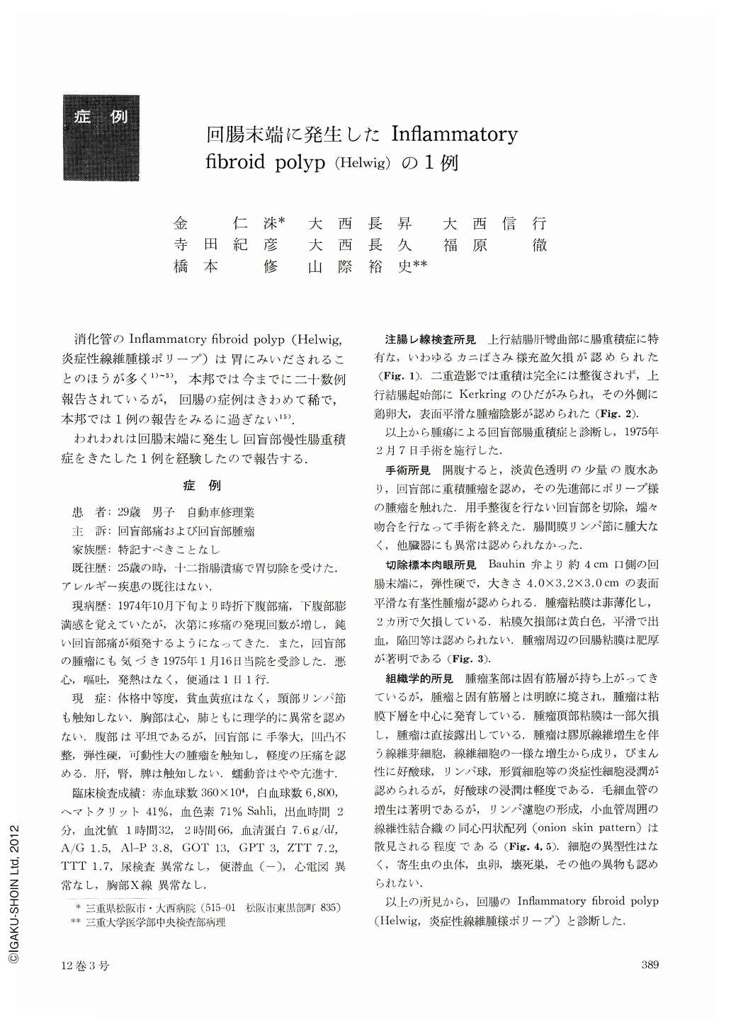
消化管のInflammatory fibroid polyp(Helwig,炎症性線維腫様ポリープ)は胃にみいだされることのほうが多く1)~5),本邦では今までに二十数例報告されているが,回腸の症例はきわめて稀で,本邦では1例の報告をみるに過ぎない15).
われわれは回腸末端に発生し回盲部慢性腸重積症をきたした1例を経験したので報告する.
A 29-year-old man visited our hospital, complaining of intermittent ileocecal pain for 2 months and an ileocecal mass. His past history was unremarkable except for duodenal ulcer for which gastrectomy had been done at our hospital at the age of 25 years. There was no significant family history. On admission, the abdomen was slightly distended, and bowel sounds were mildly accentuated. In the ileocecal region, a mobile, elastic hard, uneven, fist-sized mass was palpated. Barium enema showed a filling defect at the end of barium column. Under the diagnosis of chronic ileocecal intussusception caused by a tumor of the terminal ileum, ileo-colostomy was performed on February 7,1975. In the surgical specimen, a pedunculated polyp, measuring 4.0×3.2×3.0 cm, was situated at the ileum about 4 cm oral from the ileocecal valve. It was elastic, firm in consistency, and its surface was smooth and attenuated. The tip was ulcerated in two parts. Microscopically, the lesion was in the submucosa and consisted of hyperplasia of mesenchymal tissur, i.e, irregularly distributed fibroblasts, loosely arranged collagenous fibers, and diffusely infiltrated eosinophils, lymphocytes and plasma cells. The onion skin pattern of loosely arranged, fibrillar connective tissue about the wall of blood vessels and rudimentary lymph follicles were found here and there. There were neither parasitic bodies nor any other foreign bodies in the lesion. No malignant atypical cells were seen.
According to these findings this case was diagnosed as inflammatory fibroid polyp of the ileum. This is the second such case in Japan.
Copyright © 1977, Igaku-Shoin Ltd. All rights reserved.