Japanese
English
- 有料閲覧
PPV購入案内
1,320円 (1,200円+税10%) こちらは、700ページから704ページの文献です。購入には医書.jp本会員登録が必要です。
会員登録完了後に改めて上記「購入サイトへ」を選択してください。
または「購入サイトへ」選択後に「購入ログイン」、「新規会員登録」の順に進んでください。
会員登録について詳しくはをご確認ください。
- Abstract 文献概要
- 1ページ目 Look Inside
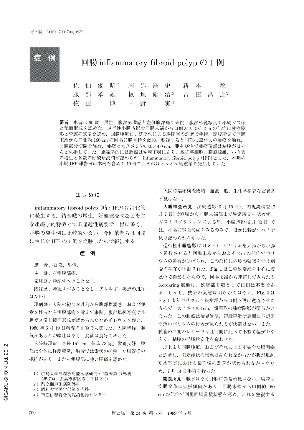
要旨 患者は60歳,男性.腹部膨満感と左側腹部痛で来院,腹部単純写真で小腸ガス像と鏡面形成を認めた.逆行性小腸造影で回腸末端から口側おおよそ2mの部位に腫瘤陰影と管腔の狭窄を認め,回腸腫瘍およびそれによる腸閉塞の診断で手術.開腹所見で回腸末端から口側約160cmの回腸に腸重積を認め,整復すると同部に鶏卵大の腫瘤を触れ,回腸部分切除を施行.腫瘤は大きさ3.5×4.0×4.Ocm,亜有茎性で腫瘤頂部は粘膜がほとんど欠損していた.組織学的には腫瘤は粘膜下層にあり,線維芽細胞,膠原線維,小血管の増生と多数の好酸球浸潤が認められ,inflammatory fibroid polyp(IFP)とした.本邦の小腸IFP報告例は本例を含めて19例で,そのほとんどが腸重積で発症していた.
A 60-year-old man was admitted to our hospital complaining of abdominal fullness and left side abdominal pain. On the abdominal plain x-ray film, bowel gas and fluid level indicated obstruction of the small intestine. Retrograde small intestinal radiography revealed a tumor-like shadow and stenosis in the ileum approximately 2 meter from the ileocaecal valve (Figs. 1, 2). It was diagnosed as a small intestinal obstruction due to a tumor. Subsequently, an operation was performed. During laparotomy, the intussusception due to the tumor was observed in the ileum, and partial ileotomy was carried out. The resected specimen showed a polypoid tumor measuring 4.0×3.5×4.0 cm, and the surface of the tumor was erosive (Fig. 3). Histologic examination revealed a tumor in the submucosal layer, consisting of hyperplasia of fibroblasts, collagen fibers and blood vessels, with eosinophilic infiltration. From these findings, the tumor was diagnosed as being an inflammatory fibroid polyp (IFP) (Figs. 5), 6).
Copyright © 1989, Igaku-Shoin Ltd. All rights reserved.