Japanese
English
- 有料閲覧
PPV購入案内
1,320円 (1,200円+税10%) こちらは、171ページから178ページの文献です。購入には医書.jp本会員登録が必要です。
会員登録完了後に改めて上記「購入サイトへ」を選択してください。
または「購入サイトへ」選択後に「購入ログイン」、「新規会員登録」の順に進んでください。
会員登録について詳しくはをご確認ください。
- Abstract 文献概要
- 1ページ目 Look Inside
- サイト内被引用 Cited by
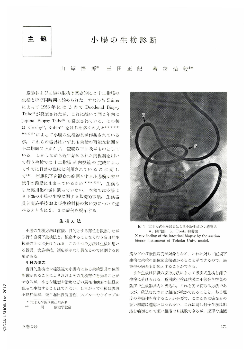
空腸および回腸の生検は歴史的には十二指腸の生検とほぼ同時期に始められた.すなわちShinerによって1956年にはじめてDuodenal Biopsy Tube1)が発表されたが,これに続いて同じ年内にJejunal Biopsy Tube2)も発表されている.その後はCrosby3),Rubin4)をはじめ多くの人々5)6)7)8)9)10)11)12)によって小腸の生検器具が作製されているが,これらの器具はいずれも生検の可能な範囲を十二指腸に止まらず,空腸以下に及ぶものとしている.しかしながら近年始められた内視鏡を用いて行う生検では十二指腸が内視鏡の完成によってすでに日常の臨床に利用されているのに対して13),空腸以下を観察の範囲とする小腸鏡は未だ試作の段階に止まっているため14)15)16)17),生検もまた実用化の域に到っていない.本稿では空腸より下部の小腸の生検に関する基礎的事項,生検器具と実施手技および生検材料の扱い方について述べるとともに2,3の症例を提示する.
Biopsy of the small intestine is performed either “blindly” or under direct vision. Blind biopsy is indicated for diffuse mucosal alterations and direct vision biopsy in addition also for localized lesions. Blind biopsy is a suctional procedure for which wire, air or water pressure is employed in operating biopsy forceps. Endoscopy for direct vision biopsy is now being exploited for obtaining still better results. There are three ways of inserting endoscopy into the digestive tract: peroral, per anum and guiding with a string. Specimens obtained by our biopsy instrument (Tohokudai type) measured 3~6 mm in diameter, mostly containing tissue of the lamina propria. Biopsy particles taken out by biopsy under direct vision by means of endoscope were 1~2 mm in diameter. The size of a given specimen was found to be greatly related to that of the forceps. Biopsy is a safe procedure with few complications.
Macroscopic observation of the mucosa of the small intestine is of great help in appraising the state of the villi. By observing, we were able to divide the normal state of the villi into three groups. Microscopic sections must be cut vertical to the mucosal surface for correct histologic diagnosis of changes in the villi. Pathologic changes should by all means be discriminated from artefacts. Deformity and atrophy of the villi are changes characteristic to the mucosal lesions of the small intestine. As a basis of correct diagnosis, our results of the measurement of the villi in each segment of the small intestine are presented here. Cases are also described, showing various pathologic changes and functional examinations related to them.
Copyright © 1974, Igaku-Shoin Ltd. All rights reserved.