Japanese
English
- 有料閲覧
PPV購入案内
1,320円 (1,200円+税10%) こちらは、911ページから916ページの文献です。購入には医書.jp本会員登録が必要です。
会員登録完了後に改めて上記「購入サイトへ」を選択してください。
または「購入サイトへ」選択後に「購入ログイン」、「新規会員登録」の順に進んでください。
会員登録について詳しくはをご確認ください。
- Abstract 文献概要
- 1ページ目 Look Inside
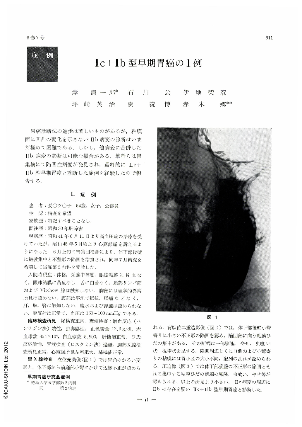
胃癌診断法の進歩は著しいものがあるが,粘膜面に凹凸の変化を示さないⅡb病変の診断はいまだ極めて困難である.しかし,他病変に合併したⅡb病変の診断は可能な場合がある.筆者らは胃集検にて陥凹性病変が発見され,最終的にⅡc+Ⅱb型早期胃癌と診断した症例を経験したので報告する.
A 57-year-old woman was referred for thorough examination because rugal convergency and irregularshaped depression on the posterior wall in the lower body had been pointed out at a gastric mass survey. The lesion was visualized by the subsequent x-ray on the corresponding site slightly on the lesser curvature side, with the abruptly ceased mucosal folds converging toward it altered at the tips: either raised, fallen away, looking as if worm-eaten, or showing club-like swelling. The mucosal surface around the lesion was slightly uneven, and the areae gastricae were varying in size or in disorderly arrangement. Endoscopy also revealed the irregular-shaped depression on the posterior wall of the lower body with an inlet-like engorged protrusion in its center. The ceased tips of the mucosal folds centering on the lesion were either tapering, looking as if vermiculated, or knobby. The mucosal surface around the concavity was slightly discolored with loss of luster. It was duly diagnosed as Ⅱc+Ⅱb type early cancer.
In the resected stomach the lesion was seen as a long, shallow depression, measuring 1.2×0.8cm, located on the posterior wall a little above the incisura. There was a whitish, discolored area on the mucosa around the lesion and it had transverse spread. The border between it and the normal mucosa was not distinct, nor was there any difference in the level of the mucosal surface.
Pathologically, it was carcinoma simplex mucocellulare mostly of intramucosal spread with the depressed lesion as its center. The muscularis mucosae was partly broken off and cicatricized, with fibrosis in the submucosa, within which was seen the only submucosal cancer infiltration consisting of several cancer cells. On the other hand, intramucosal involvement was far more extrensive than the area of depression. Though irregular and discontinuous, its extent seemed to correspond approximately to the whole discolored area.
The case here described illustrates the necessity of careful diagnosis even of a seemingly Ⅱc lesion with due observation of the areas around the lesion, especially of their nature, discoloration or loss of luster.
Copyright © 1971, Igaku-Shoin Ltd. All rights reserved.