Japanese
English
- 有料閲覧
- Abstract 文献概要
- 1ページ目 Look Inside
はじめに
胃壁内好酸球性肉芽腫はJaegerにより1932年初めて報告され,その後わが国においても多くの人により発表されている.筆者らは最近,胃にポリープ様腫瘤を呈し,Ⅰ型早期胃癌を思わせた好酸球性肉芽腫の1例を経験したので報告する.
This paper deals with a case of eosinophilic granuloma resembling early gastric cancer of type I because of its polypoid appearance.
Case: a 63-year-old man desiring thorough examination of his stomach.
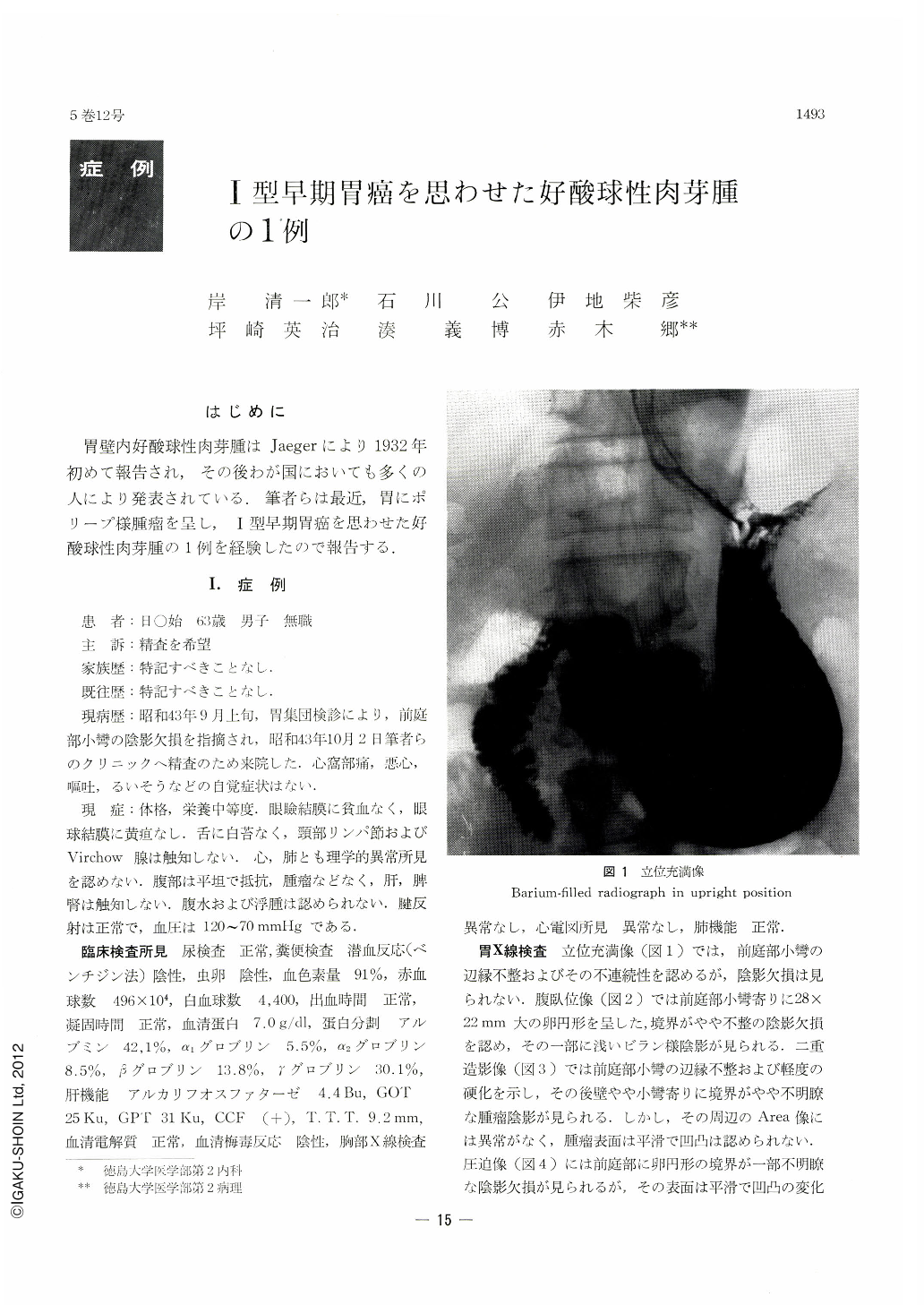
At x-ray examination of the stomach, irregular and uneven contour of the lesser curvature was seen in an upright, barium-filled picture, together with an oval shadow defect in the antrum as visualized in prone position and also by compression. The shadow defect with partly indistinct margin was of smooth surface. No bridging fold was recognized. Endoscopy also revealed a fairly large oval protrusion on the lesser curvature of the antrum. Its surface was slightly uneven, and erosions and white exudate were seen on a greater part of it. By these findings, submucosal tumor, polyp or atypical epithelium was at first taken into account, but finally early gastric cancer of type I became most suspicious. Operation was accordingly performed. Macroscopically, a globular polyp measuring 20×17×16 mm, with a stalk 7mm in diameter, was seen on the lesser curvature of the antrum. It was of elasitic soft consistency. Its surface, dark red and smooth, showed normal highlights. No particular change was noticed on other parts of the mucosa. Histologically, the polyp consisted mainly of hyperplasia of the connective tissues and diffuse infiltration of eosinophilic cells, accompanied with formation of lymph follicles.
The absence of any parasitic larva body, ova or necrotic nest shows that this tumor is not a parasitic granuloma due to Anisakis, but an inflam-matory fibroid polyp, corresponding to “localized eosinophilic granuloma” as defined by McCune.

Copyright © 1970, Igaku-Shoin Ltd. All rights reserved.


