Japanese
English
- 有料閲覧
- Abstract 文献概要
- 1ページ目 Look Inside
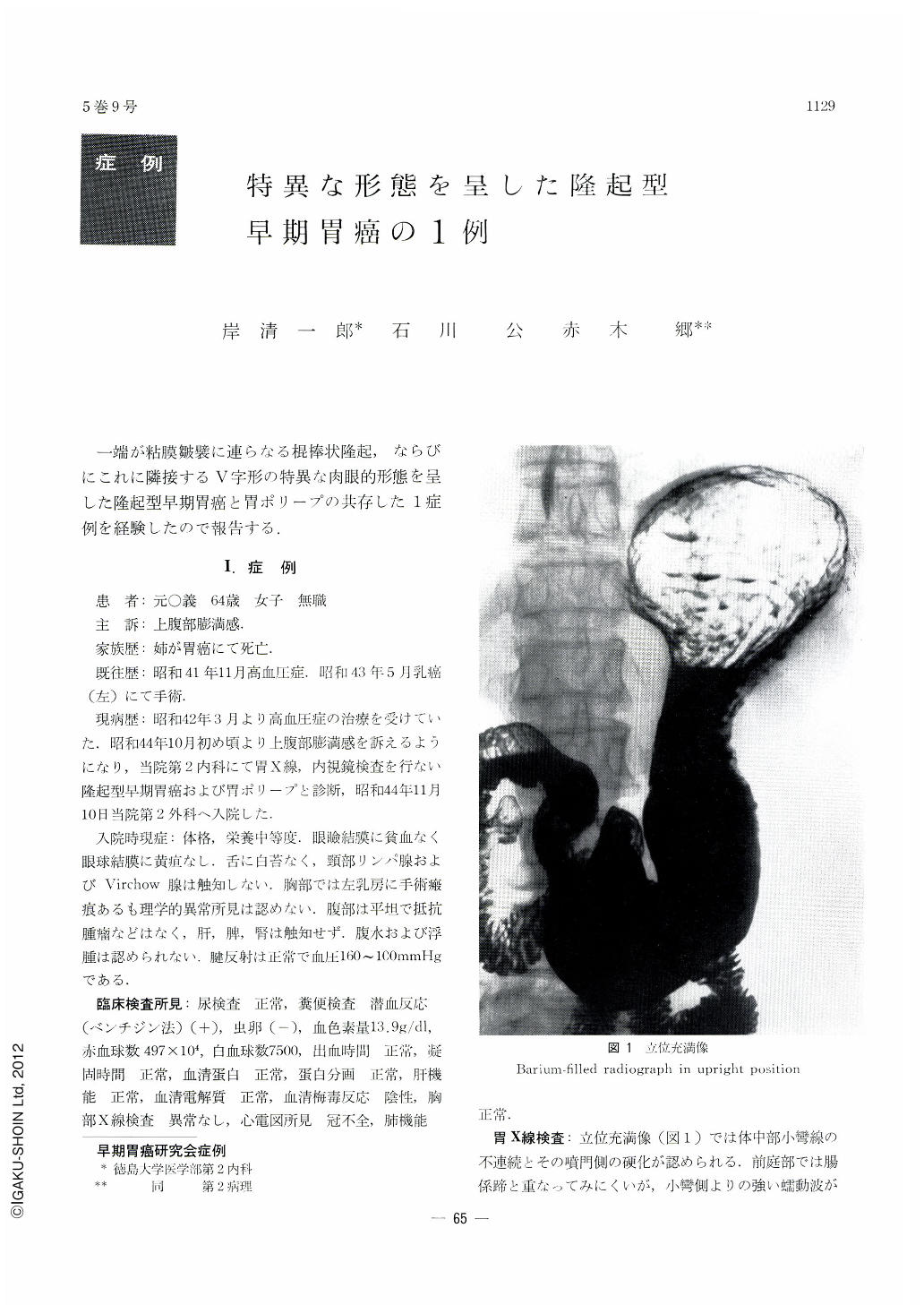
一端が粘膜皺襞に連らなる棍捧状隆起,ならびにこれに隣接するV字形の特異な肉眼的形態を呈した隆起型早期胃癌と胃ポリープの共存した1症例を経験したので報告する.
1.症例
患者:元○義 64歳 女子 無職
主訴:上腹部膨満感
家族歴:姉が胃癌にて死亡.
既往歴:昭和41年11月高血圧症.昭和43年5月乳癌(左)にて手術.
現病歴:昭和42年3月より高血圧症の治療を受けていた.昭和44年10月初め頃より上腹部膨満感を訴えるようになり,当院第2内科にて胃X線,内視鏡検査を行ない隆起型早期胃癌および胃ポリープと診断,昭和44年ll月10日当院第2外科へ入院した.
Early gastric cancer of type I having a peculiar gross shape associated with benign polyps was found in a 64-year-old female complaining of feeling of fullness in the upper abdomen. The cancer lesion located in the corpus looked at a first glance in x-ray examination like a submucosal tumor because the protruding lesion continuous with a mucosal fold appeared like a bridging fold. On further observation, however, an apparent border was recognized between the mucosal fold and the tumor so that the latter was regarded as a mucosal protrusion. Endoscopy revealed slight unevenness and discoloration on the surface of the elevation with partial erosion and bleeding. The diagnosis of protruding type of early cancer was thus arrived at. The other protuberance in the antrum was undoubtedly confirmed both by x-ray and endoscopy as a benign polyp. Gross as well as histological picture of the resected specimen showed a clublike protrusion measuring 53×10×5 mm in an area extending from the lesser curvature of the corpus away to the posterior wall. Adjacent to it, there was another elevation shaped like the letter V, measuring 30×22×5 mm, with its one end fused into a rnucosal fold running toward the posterior wall. Between the two protrusions was seen a thin groove-like boundary. The surface of the latter protrusion was granular with a distinct variance of hue between it and the continuing mucosal fold. Well differentiated adenocarcinoma tubulare with submucosalinfiltration was recognized in the area nearly corresponding with the extent of the two lesions. The other two protruding lesions in the antrum were both adenomatous polyps.
Because of their peculiar shapes, two protrusions in the corpus should be diffentiated from non-epithelial tumor; they must be confirmed by biospy.

Copyright © 1970, Igaku-Shoin Ltd. All rights reserved.


