Japanese
English
- 有料閲覧
- Abstract 文献概要
- 1ページ目 Look Inside
症例
患 者:I. U. 47歳,女.
主 訴:心窩部痛.
X線検査:46年3月4日.
内視鏡検査:46年3月5日.
手 術:46年3月14日.
X線所見
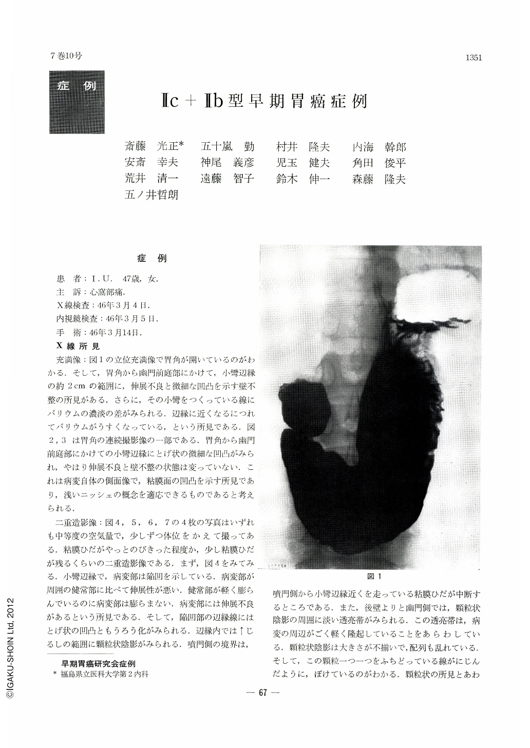
充満像:図1の立位充満像で胃角が開いているのがわかる.そして,胃角から幽門前庭部にかけて,小彎辺縁の約2cmの範囲に,伸展不良と微細な凹凸を示す壁不整の所見がある.さらに,その小彎をつくっている線にバリウムの濃淡の差がみられる.辺縁に近くなるにつれてバリウムがうすくなっている,という所見である.図2,3は胃角の連続撮影像の一部である.胃角から幽門前庭部にかけての小彎辺縁にとげ状の微細な凹凸がみられ,やはり伸展不良と壁不整の状態は変っていない.これは病変自体の側面像で,粘膜面の凹凸を示す所見であり,浅いニッシェの概念を適応できるものであると考えられる.
The patient is a 47-year-old woman with a diagnosis of II c subtype early gastric cancer, measuring 22×20 cm, located on the posterior wall from the level of the angle down to the pyloric antrum. Depth of invasion was m. Histologic type : signet ring cell carcinoma, intermixed partially with well-differentiated adenocarcinoma.
Gross findings of the resected stomach : There were few macroscopic changes. The area of cancer was almost of the same height as the surrounding normal mucosa. In the center of the lesion and in its posterior wall side was the granular change characterized by difference in size of granules with their outline presenting worm-eaten picture. Otherwise the change was unnoticiable.
X-ray findings : Serial radiographs in double contrast, with the air so proportioned that the mucosal folds were not too flattened out, revealed the lesion as a depressed area along the margin of the lesser curvature. Macroscopically, there had been scarcely any change of height between this area and the other adjoining mucosa. The lesion pictured in x-ray was encircled in its whole circumference by a thin strip of radiolucency. These findings were interpreted as arising out of difference in distensibility between the involved and normal areas. The fact that the lesion was more distinct in x-ray than in gross observation seems to show that x-ray can demonstrate lessened distensibility of the gastric wall or its rigidity otherwise difficult to find out. When serial exposures are taken in double contrast with well proportioned air within the stomach, slight cancerous changes scarcely visible to the naked eye seem to be now within reach of x-ray diagnosis.

Copyright © 1972, Igaku-Shoin Ltd. All rights reserved.


