Japanese
English
- 有料閲覧
- Abstract 文献概要
- 1ページ目 Look Inside
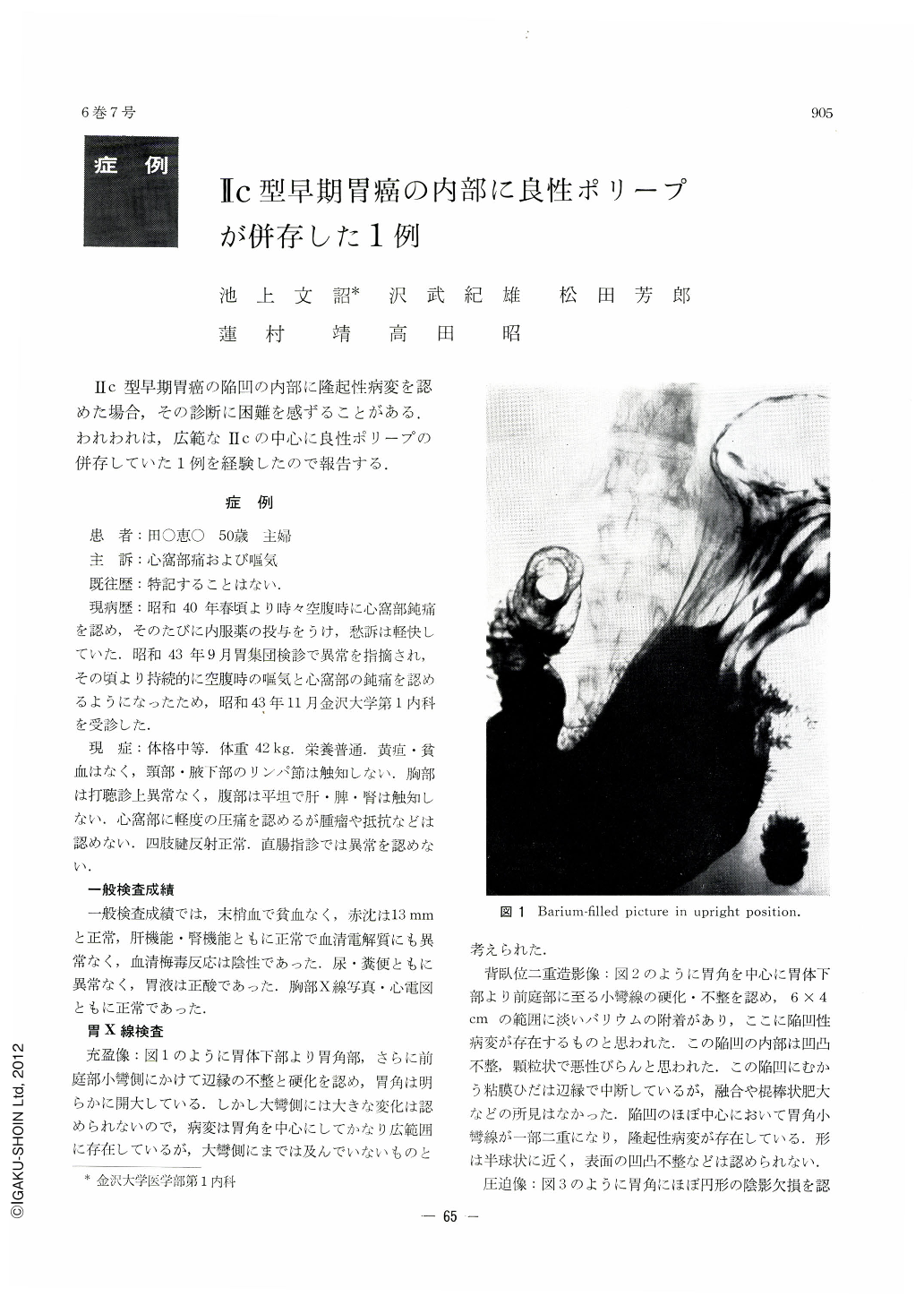
Ⅱc型早期胃癌の陥凹の内部に隆起性病変を認めた場合,その診断に困難を感ずることがある.われわれは,広範なⅡcの中心に良性ポリープの併存していた1例を経験したので報告する.
A fifty-year-old housewife, who complained of upper abdominal pain, consulted our clinic for close examination of the stomach. When first seen, she did not show any abnormal physical findings, except for tenderness on the epigastric region, and various laboratory tests were also within normal ranges.
On the X-ray examination of the stomach, filling picture of the lesser curvature between the corpus and the antrum was irregular and rigid. By the double contrast or compression study, a relatively wide depressed area with uneven surface was found between the lower part of the corpus and the antrum. The mucosal folds were interrupted at the depressed area. In addition, a small polypoid lesion was observed at the center of the depressed area. By gastrofiberscopic examination, a small polypoid lesion was seen on the angulus of the stomach. Mucosa surrounding the polypoid lesion seemed to be depressed irregularly and its surface was uneven and coated with a white fur. These findings suggested the existence of early gastic cancer of type Ⅱc, but the nature of the small polypoid lesion was obscure.
Resected specimen revealed a relatively wide depressed area (7×7 cm) through the corpus to the antrum and the gastric folds were broken down around the depressed area. In the center of the depressed lesion was a small polyp (8×4×6 mm) Histological diagnosis of the depressed area was carcinoma solidum simplex mucocellulare medullare and the cancer cells localized through only the mucosa indicating early gastric cancer. A small polyp was diagnosed as benign adenomatous polyp. No cancer cell was seen at the tip of the polypoid lesion.
These results indicated that early gastric cancer of Ⅱc type had infilterated into the adenomatous polyp at the angulus, which might have developed independently to cancer.

Copyright © 1971, Igaku-Shoin Ltd. All rights reserved.


