Japanese
English
- 有料閲覧
- Abstract 文献概要
- 1ページ目 Look Inside
- サイト内被引用 Cited by
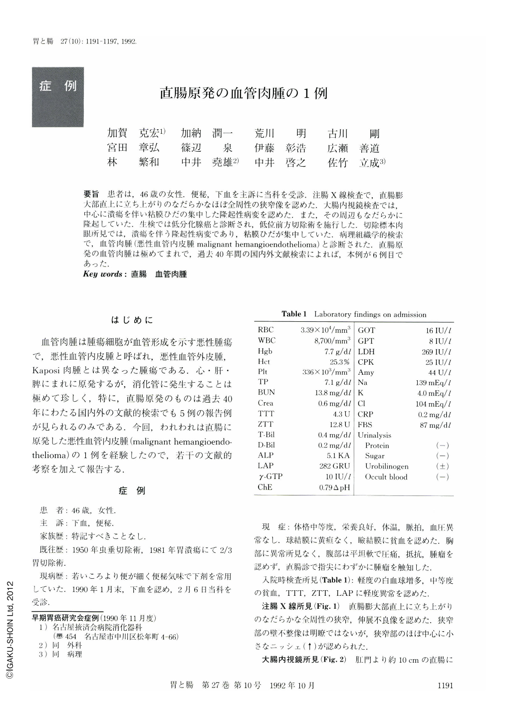
要旨 患者は,46歳の女性.便秘,下血を主訴に当科を受診.注腸X線検査で,直腸膨大部直上に立ち上がりのなだらかなほぼ全周性の狭窄像を認めた.大腸内視鏡検査では,中心に潰瘍を伴い粘膜ひだの集中した隆起性病変を認めた.また,その周辺もなだらかに隆起していた.生検では低分化腺癌と診断され,低位前方切除術を施行した.切除標本肉眼所見では,潰瘍を伴う隆起性病変であり,粘膜ひだが集中していた.病理組織学的検索で,血管肉腫(悪性血管内皮腫malignant hemangioendothelioma)と診断された.直腸原発の血管肉腫は極めてまれで,過去40年間の国内外文献検索によれば,本例が6例目であった.
A 46-year-old woman was admitted to our hospital with the chief complaint of constipation and hematochezia. A barium enema showed stenosis with a small niche (↑) and a smooth margin in the rectum (Fig. 1). Colonoscopic examination showed a smoothly elevated lesion and an ulcer with an irregular margin and converging folds (Fig. 2).
Pelvic CT scan revealed a thick posterior wall of the rectum with positive enhancement and enlarged regional lymph nodes (Fig. 3). Histological examination of a punch biopsy specimen showed poorly differentiated adenocarcinoma (Fig. 4). An operation was performed. There was a protruding lesion with an irregular ulcer and fold convergency (Fig. 5a). A cut section of the specimen showed clearly demarcated layers of the rectum. The muscularis was dark and thickened secondary to tumor cell infiltration (Fig. 5b). Microscopic examination showed infiltration of tumor cells into the lamina propria and subserosa (Fig. 6a). Some tumor cells formed tubular structures containing red blood cells (Fig. 6b) and others were solid (Fig. 6c). The pathologic diagnosis was malignant hemangioendothelioma, based on finding of proliferating tumor cells within the reticulin sheath (Fig. 6d). Almost all resected lymph nodes revealed metastasis (Fig. 6e). Malignant hemangioendothelioma comprises 1% of all sarcomas and is rare in the rectum. We could find only 5 such cases in the literature of the past 40 years.

Copyright © 1992, Igaku-Shoin Ltd. All rights reserved.


