Japanese
English
- 有料閲覧
PPV購入案内
1,320円 (1,200円+税10%) こちらは、1009ページから1020ページの文献です。購入には医書.jp本会員登録が必要です。
会員登録完了後に改めて上記「購入サイトへ」を選択してください。
または「購入サイトへ」選択後に「購入ログイン」、「新規会員登録」の順に進んでください。
会員登録について詳しくはをご確認ください。
- Abstract 文献概要
- 1ページ目 Look Inside
- サイト内被引用 Cited by
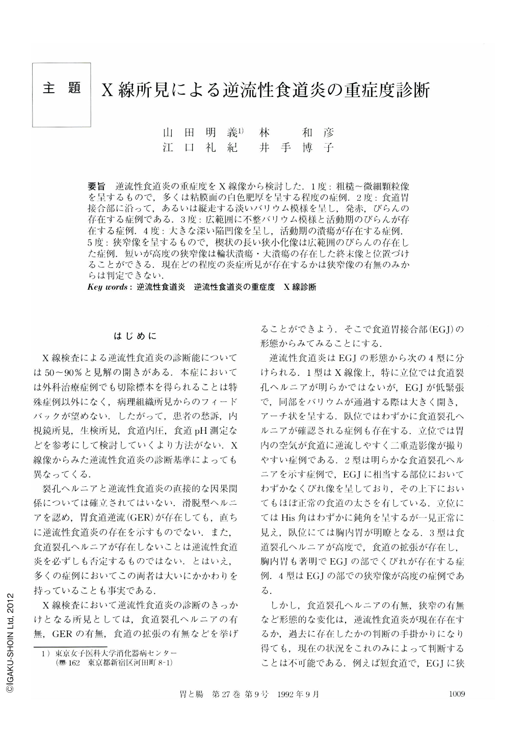
要旨 逆流性食道炎の重症度をX線像から検討した.1度:粗糙~微細顆粒像を呈するもので,多くは粘膜面の白色肥厚を呈する程度の症例.2度:食道胃接合部に沿って,あるいは縦走する淡いバリウム模様を呈し,発赤,びらんの存在する症例である.3度:広範囲に不整バリウム模様と活動期のびらんが存在する症例.4度:大きな深い陥凹像を呈し,活動期の潰瘍が存在する症例.5度:狭窄像を呈するもので,楔状の長い狭小化像は広範囲のびらんの存在した症例.短いが高度の狭窄像は輪状潰瘍・大潰瘍の存在した終末像と位置づけることができる.現在どの程度の炎症所見が存在するかは狭窄像の有無のみからは判定できない.
We classified severity of reflux esophagitis on barium swallowing examination as follows.
Grade 1: Hypertrophic mucosa of esophagus showing rough and fine granular pattern.
Grade 2: Reddish and/or erosive mucosal lesion showing longitudinal linear ulcerative pattern along EG junction.
Grade 3: Extensive active erosion showing an extensive irregular surface.
Grade 4: Active ulcer with deep excavation.
Grade 5: Stenotic lesion. An extensive erosive lesion resulted in a long wedge-shaped long stenosis. A large, annular ulcer caused a well-demarcated but severe stenotic lesion.
The severity of stenosis does not indicate the extent of inflammation.
Copyright © 1992, Igaku-Shoin Ltd. All rights reserved.